


Báo cáo khoa học: "A rare tumoral combination, synchronous lung adenocarcinoma and mantle cell lymphoma of the pleura"
lượt xem 4
download
 Download
Vui lòng tải xuống để xem tài liệu đầy đủ
Download
Vui lòng tải xuống để xem tài liệu đầy đủ
Tuyển tập báo cáo các nghiên cứu khoa học quốc tế ngành y học dành cho các bạn tham khảo đề tài: A rare tumoral combination, synchronous lung adenocarcinoma and mantle cell lymphoma of the pleura
Bình luận(0) Đăng nhập để gửi bình luận!
Nội dung Text: Báo cáo khoa học: "A rare tumoral combination, synchronous lung adenocarcinoma and mantle cell lymphoma of the pleura"
- World Journal of Surgical Oncology BioMed Central Open Access Case report A rare tumoral combination, synchronous lung adenocarcinoma and mantle cell lymphoma of the pleura Dimitrios Hatzibougias1, Mattheos Bobos1, Georgia Karayannopoulou1, Georgios Karkavelas1, Georgios T Karapanagiotidis2, Christophoros N Foroulis*2 and Ioannis Kostopoulos1 Address: 1Aristotle University of Thessaloniki Medical School, Department of Pathology, Thessaloniki, Greece and 2Aristotle University of Thessaloniki Medical School, AHEPA University Hospital, Department of Cardio-Thoracic Surgery, Thessaloniki, Greece Email: Dimitrios Hatzibougias - Dhbugias@yahoo.gr; Mattheos Bobos - mbobos@auth.gr; Georgia Karayannopoulou - karayan@med.auth.gr; Georgios Karkavelas - gkarkav@med.auth.gr; Georgios T Karapanagiotidis - karapang7@hotmail.com; Christophoros N Foroulis* - cforoulis@otenet.gr; Ioannis Kostopoulos - kostop@med.auth.gr * Corresponding author Published: 29 December 2008 Received: 29 July 2008 Accepted: 29 December 2008 World Journal of Surgical Oncology 2008, 6:137 doi:10.1186/1477-7819-6-137 This article is available from: http://www.wjso.com/content/6/1/137 © 2008 Hatzibougias et al; licensee BioMed Central Ltd. This is an Open Access article distributed under the terms of the Creative Commons Attribution License (http://creativecommons.org/licenses/by/2.0), which permits unrestricted use, distribution, and reproduction in any medium, provided the original work is properly cited. Abstract Background: Coexistence of adenocarcinoma and mantle cell lymphoma in the same or different anatomical sites is extremely rare. We present a case of incidental discovery of primary lung adenocarcinoma and mantle cell lymphoma involving the pleura, during an axillary thoracotomy performed for a benign condition. Case presentation: A 73-year old male underwent bullectomy and apical pleurectomy for persistent pneumothorax. A bulla of the lung apex was resected en bloc with a scar-like lesion of the lung, which was located in proximity with the bulla origin, by a wide wedge resection. Histologic examination of the stripped-off parietal pleura and of the bullectomy specimen revealed the synchronous occurrence of two distinct neoplasms, a lymphoma infiltrating the pleura and a primary, early lung adenocarcinoma. Immunohistochemical and fluorescence in situ hybridization assays were performed. The morphologic, immunophenotypic and genetic findings supported the diagnosis of primary lung adenocarcinoma (papillary subtype) coexisting with a non-Hodgkin, B-cell lineage, mantle cell lymphoma involving both, visceral and parietal pleura and without mediastinal lymph node involvement. The neoplastic lymphoid cells showed the characteristic immunophenotype of mantle cell lymphoma and the translocation t(11;14). The patient received 6 cycles of chemotherapy, while pulmonary function tests precluded further pulmonary parenchyma resection (lobectomy) for his adenocarcinoma. The patient is alive and without clinical and radiological findings of local recurrence or distant relapse from both tumors 14 months later. Conclusion: This is the first reported case of a rare tumoral combination involving simultaneously lung and pleura, emphasizing at the incidental discovery of the two coexisting neoplasms during a procedure performed for a benign condition. Any tissue specimen resected during operations performed for non-tumoral conditions should be routinely sent for pathologic examination. Page 1 of 5 (page number not for citation purposes)
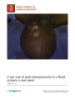
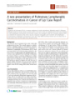
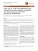
- World Journal of Surgical Oncology 2008, 6:137 http://www.wjso.com/content/6/1/137 Background Case presentation Recent epidemiologic evidence suggest that lung cancer is A 73-year old man, heavy smoker, with history of chronic the leading cause of cancer mortality in both sexes and is obstructive pulmonary disease, coronary artery disease distinguished according to histopathologic features in two and HBV infection was admitted to a district hospital large categories, the small cell lung carcinoma (SCLC) and because of dyspnea and fever (38–38.5°C). Clinical and the non-small cell lung carcinoma (NSCLC) [1]. The most radiologic findings revealed spontaneous pneumothorax common type of the latter is lung adenocarcinoma which on the right side, which was initially managed by chest is usually presented as single or multiple poorly circum- tube drainage. Due to persistent for more than 10 days air scribed peripheral lung lesions [2]. Papillary adenocarci- leak through the chest tube, he was referred to the depart- noma is a rare histopathologic subtype of lung ment of Cardiothoracic Surgery at AHEPA University Hos- adenocarcinoma which is characterized by predominantly pital. CT scan imaging of the thorax showed diffuse papillary structures that replaces the underlying lung emphysema, surgical subcutaneous emphysema on the parenchyma and has been stated to be associated with right side as the result of previous chest tube drainage, a poorer prognosis [3,4]. scar-like lesion that was located in the periphery of the posterior segment of the right upper lobe (Fig. 1a) and Mantle cell lymphoma (MCL) is a distinct type of B-cell thickening of parietal pleura adjacent to the lung scar (Fig. non-Hodgkin lymphoma characterized by 1b). Because of the prolonged air leak, the patient under- t(11;14)(q13;q32) and Cyclin D1 over-expression, com- went apical bullectomy and apical parietal pleura resec- prising from 3% to 10% of all non-Hodgkin's lymphomas tion to achieve pleurodesis through an axillary [5,6]. The affected patients are mainly middle-aged or thoracotomy. The scar-like lesion of the lung was resected older and they are often presented with advanced stage en bloc with the bullectomy specimen by a wide wedge disease (Stages III-IV), frequently involving multiple resection of lung parenchyma. extranodal sites [7]. Pathological findings and immunohistochemistry Synchronous occurrence of lung adenocarcinoma and On macroscopic examination of the bullectomy speci- malignant lymphoma of the pleura is not reported until men, a solid yellowish nodular lesion measuring up to 1.6 today and we report the unique case of a lung adenocarci- cm was found. Close to that lesion many cystic spaces noma coexisting with a mantle cell lymphoma of the were found. pleura, which were incidentally discovered during an operation for pneumothorax. The histologic examination of hematoxylin-eosin-stained sections revealed an adenocarcinoma composed of atypi- Figure 1 Preoperative chest CT scan Preoperative chest CT scan. (A) Diffuse emphysematous changes of the right upper lobe, surgical subcutaneous emphy- sema and a scar-like lesion located in the periphery of the posterior segment of the right upper lobe (arrow). (B) Thickening of the parietal pleura in proximity to the scar-like lesion (arrow). Page 2 of 5 (page number not for citation purposes)
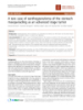
- World Journal of Surgical Oncology 2008, 6:137 http://www.wjso.com/content/6/1/137 Figure 2 Lung adenocarcinoma with micropapillary pattern (A, E & H) Lung adenocarcinoma with micropapillary pattern (A, E & H). Neoplastic ducts showing strong nuclear staining for TTF-1 (B) and cytokeratin 7 (C). Mantle cell lymphoma with diffuse infiltration of the pleura (D, E & H). Lymphomatous cells, positive for CD5 (E) and cyclin D1 (F). FISH method using the LSI IGH/CCND1 dual color, dual fusion translocation probe: nucleus of neoplastic lymphoid cell with t(11;14) displaying the signal pattern 2 yellow (fusion translocation signals), 2 red (CCND1 gene) and 2 green (IGH gene) (G). Page 3 of 5 (page number not for citation purposes)
- World Journal of Surgical Oncology 2008, 6:137 http://www.wjso.com/content/6/1/137 cal adenoid formations showing intraluminal papillary Discussion projections, some of them detached (Fig. 2a). The tumor Synchronous occurrence of lung adenocarcinoma and cells were tall-cylindrical, with pale eosinophilic cyto- mantle cell lymphoma of the pleura is not reported in the plasm and polymorphic nuclei with or without promi- medical literature until today. Extensive search of the lit- nent nucleoli. The mitotic activity was low (< 2 mitoses erature revealed few cases with coexistence of different per 10 HPF). There were also foci of necrosis, hyalinosis types of lung carcinomas and malignant lymphomas. and ossification. Immunohistochemical stains showed Chanel et al. described a synchronous pulmonary adeno- the following tumor-cell immunophenotype: TTF-1(+) carcinoma and extranodal marginal zone lymphoma of (Novocastra, UK), CK7(+) (Dako, DK), CK20(+) (Dako), MALT type [8]. Rothenburger et al. reported a non-Hodg- EGFR(+) (Zymed, USA), p53(+) (Dako), Cyclin D1(+) kin's lymphoma coexisting with a NSCLC, whereas Rubi- (Spring, USA), Calretinin(-) (Dako) (Fig. 2b, 2c). The ales et al. described the synchronous occurrence of a adjacent lung parenchyma showed serious emphysematic small-cell lung cancer and a Hodgkin lymphoma [9,10]. changes. Coexistence of adenocarcinoma and mantle cell lym- The visceral pleura proximal to the adenocarcinoma and phoma in other anatomical sites, such as the large bowel, the separately sent to the laboratory specimen of stripped- has been reported in the past. In total we found four cases off parietal pleura tissue showed a band-like diffuse infil- concerning colonic involvement. Hopster et al, described tration from neoplastic lymphoid cells (Fig. 2d). The lym- 2 foci of colonic adenocarcinoma associated with MCL phoid cells were medium-sized, with rounded or angular [11]. Kanehira et al., presented also 2 cases of invasive ade- nuclei and with one or more indistinct nucleoli. Focally nocarcinoma of the colon coexisting with early MCL [12]. there was a nodular pattern of growth. The immunophe- The fourth case reported by Padmanabhan et al, con- notype of the lymphoma cells was the following: cerned the synchronous presence of adenocarcinoma CD20(+) (Dako), CD45RA(+) (Dako), CD5(+) (Novo- located in the cecum and MCL involving the colon, the castra), Cyclin D1(+) (Spring), CD45RO(-) (Dako), terminal ileum and the regional lymph nodes [13]. CD3(-) (Novocastra), CD23(-) (Novocastra), EGFR(-), Another case of synchronous existence of nodal mantle Calretinin(-) (Fig. 2e, 2f). The CD23 immunostain cell lymphoma and metastatic in mediastinal lymph revealed a residual network of dendritic cells, referring to nodes small cell lung carcinoma was reported by Kampal- infiltration of preexisting germinal centers. ath et al [14]. Lung adenocarcinoma usually arises from peripheral Fluorescence in situ hybridization (FISH) Metaphase FISH analysis was also performed using EGFR/ small bronchi and may be associated with a lung scar (scar CEP7, CCND1/CEP11 dual color LSI probes and LSI IGH/ adenocarcinoma). It is composed of malignant glandular CCND1 dual color, dual fusion translocation probe (all epithelium which may vary in degree of differentiation from Abbott Molecular, USA). Gains of EGFR and from tumor to tumor. Well differentiated tumors may CCND1 genes were observed in the adenocarcinoma. On form distinct glands, while others may vary from forming the other hand, in MCL the characteristic translocation papillary structures to solid tumors without any gland for- between CCND1 gene on chromosome 11 and IGH gene mation. The prognosis depends on the histologic type, on chromosome 14 [t(11;14)] was found (Fig. 2g). In clinical stage, and the patient's performance status. A addition CCND1 gene was amplified, whereas EGFR gene micropapillary pattern is a predictor of poor prognosis was on normal range. [3,15]. The presence of this component should alert the clinician to search more carefully for clinically "silent" The morphologic, immunophenotypic and genetic find- metastases. ings support the diagnosis of primary lung adenocarci- noma (papillary subtype) coexisting with a non-Hodgkin, MCL is a neoplasm of monomorphous small to medium- B-cell lineage, mantle cell lymphoma involving both, vis- sized B lymphocytes with irregular nuclei, which resemble ceral and parietal pleura and without mediastinal lymph the cleaved cells (centrocytes) of germinal centers. Neo- node involvement. plastic transformed cells (centroblasts or immunoblasts) are absent. Tumor cells are typically CD5(+) and CD23(- ). The vast majority overexpresses Cyclin D1. Vega et al, in Follow-up The patient received 6 cycles of chemotherapy (Endoxan, their review of 34 patients with lymphoma involving the Farmorubicin and Vincristine) and his clinical status, 14 pleura that was detected by pleural biopsy, found only 1 months after the diagnosis is good, without any evidence MCL among the 34 cases [16]. The most frequent type in of local recurrence, metastatic disease or lymph node their series was diffuse large B-cell lymphoma, followed involvement, according to the follow-up CT scan imaging by follicular lymphoma. The histologic pattern of MCL of thorax, abdomen and brain. may be diffuse, nodular, or mantle zone, or a combina- Page 4 of 5 (page number not for citation purposes)
- World Journal of Surgical Oncology 2008, 6:137 http://www.wjso.com/content/6/1/137 tion of the three patterns of growth. Some reports indicate FISH assays, has drafted the manuscript and has contrib- a better prognosis for cases with a mantle zone pattern. uted to the interpretation of the results. Georgios Karkave- Despite the small size and bland appearance of these cells, las has given final approval of the version to be published. there is often more mitotic activity than in other histolog- GTK has assisted the surgeon, did the collection of the ically low-grade lymphomas. Diffuse forms and those data and have been involved in the design and drafting of which show high mitotic rates (> 20 HPF in diffuse and > the manuscript. CNF has performed the operation and 10 HPF in follicular) have a worse prognosis. has critically revised the initial draft. IK carried out pathol- ogy examination, immunohistochemistry and FISH We report a unique case of coexistence of lung adenocar- assays, has critically revised the initial draft and has given cinoma and mantle cell lymphoma of the pleura. The final approval of the version to be published patient was admitted to the hospital due to persistent air leak from chest tube after an episode of spontaneous sec- References ondary pneumothorax, with no other specific clinical 1. Parkin M, Tyczynski JE, Boffetta P, Samet J, Shields J, Caporaso N: Lung cancer epidemiology and etiology. In World Health Organ- signs, and both tumors were incidentally identified. Poor ization Classification of Tumours. Pathology and Genetics of Tumors of the results of pulmonary function tests and the coexistence of Lung, Pleura, Thymus and Heart Edited by: Travis WD, Brambilla E, Muller-Hermelink HK, Harris CC. Lyon: IARC Press; 2005:12-15. mantle cell lymphoma precluded the patient from further 2. Madri JA, Carter D: Scar cancers of the lung. Origin and signif- surgical treatment (lobectomy) for his early lung adeno- icance. Hum Pathol 1984, 15:625-631. carcinoma. 3. Amin MB, Tamboli P, Merchant SH, Ordóñez NG, Ro J, Ayala AG, Ro JY: Micropapillary component in lung adenocarcinoma: a dis- tinctive histologic feature with possible prognostic signifi- Conclusion cance. Am J Surg Pathol 2002, 26:358-364. 4. Miyoshi T, Satoh Y, Okumura S, Nakagawa K, Shirakusa T, Tsuchiya The therapeutic management of such a combination of E, Ishikawa Y: Early-stage lung adenocarcinomas with a micro- tumors requires separate consideration of their biologic papillary pattern, a distinct pathologic marker for a signifi- behavior, the performance status of each patient individ- cantly poor prognosis. Am J Surg Pathol 2002, 27:101-109. 5. Barista I, Romaguera JE, Cabanillas F: Mantle cell lymphoma. Lan- ually and the estimated morbidity related to surgery and/ cet Oncol 2001, 2:141-148. or chemo-radiotherapy. We should note that any tissue 6. de Boer CJ, Schuuring E, Dreef E, Peters G, Bartek J, Kluin PM, van resected during any non-oncologic intrathoracic proce- Krieken JH: Cyclin D1 protein analysis in the diagnosis of man- tle cell lymphoma. Blood 1995, 86:2715-2723. dure should be collected separately and sent for patho- 7. Pittaluga S, Wlodarska I, Stul MS, Thomas J, Verhoef G, Cassiman JJ, logic examination, especially in older people. Any scar Berghe H Van den, de Wolf-Peeters C: Mantle cell lymphoma. A clinicopathological study of 55 cases. Histopathology 1995, detected in the lung should also resected during an 26:17-24. intrathoracic procedure performed for benign disease, if 8. Chanel S, Burke L, Fiche M, Molina T, Lerochais JP, Icard P, Diebold J, do not require a major operation and do not add signifi- Galateau-Sallé F: Synchronous pulmonary adenocarcinoma and extranodal marginal zone/low- grade B-cell lymphoma cant risk for the patient. of MALT type. Hum Pathol 2001, 32:129-132. 9. Rothenburger M, Semik M, Hoffmeier A, Baba H, Kamanabrou D, Roos N, Schmidt C, Scheld HH: Coexistence of non-Hodgkin's Abbreviations lymphoma and non-small cell lung carcinoma: diagnosis and SCLC: squamous cell lung cancer; NSCLC: non small cell treatment. Thorac Cardiovasc Surg 2002, 50:59-61. lung cancer; MCL: mantle cell lymphoma; HBV: hepatitis 10. Rubiales AS, Martinez G, Aller JL, Roig V, del Valle ML: Synchronous diagnosis of small-cell lung cancer and Hodgkin lymphoma. B virus; CT: computed tomography; FISH: fluorescence in An Med Interna 2006, 23:301-302. situ hybridization; HPF: high power field; MALT: mucosa- 11. Hopster D, Smith PA, Nash JR, Elders K, Poston GJ: Synchronous associated lymphoid tissue. multiple lymphomatous polyposis and adenocarcinomas of the large bowel. Postgrad Med J 1995, 71:443. 12. Kanehira K, Braylan RC, Lauwers GY: Early phase of mantle cell Consent lymphoma: a report of 2 cases associated with advanced colonic adenocarcinoma. Mod Pathol 2001, 14:811-817. Written informed consent was obtained from the patient 13. Padmanabhan V, Trainer TD: Synchronous adenocarcinomas for publication of this case report and any accompanying and mantle cell lymphoma of the colon. Arch Pathol Lab Med images. A copy of the written consent is available for 2003, 127:E64-66. 14. Kampalath B, Abed N, Chitambar CR, Vantuinen P, Chakrabarty G, review by the Editor-in-Chief of this journal. Hanson G, Rao RN, Shidham VB, Chang CC: Mantle cell lym- phoma in lymph nodes with metastatic small cell carcinoma Competing interests of lung: a diagnostic and treatment dilemma. Leuk Lymphoma 2004, 45:409-414. The authors declare that they have no competing interests. 15. Tsutsumida H, Nomoto M, Goto M, Kitajima S, Kubota I, Hirotsu Y, Wakimoto J, Hollingsworth MA, Yonezawa S: A micropapillary pattern is predictive of a poor prognosis in lung adenocarci- Authors' contributions noma, and reduced surfactant apoprotein An expression in DH carried out pathology examination, immunohisto- the micropapillary pattern is an excellent indicator of a poor chemistry and drafted the manuscript. MB carried out prognosis. Mod Pathol 2007, 20:638-647. 16. Vega F, Padula A, Valbuena JR, Stancu M, Jones D, Medeiros LJ: Lym- immunohistochemistry and FISH assays and drafted the phomas involving the pleura: A clinicopathological study of manuscript. Georgia Karayannopoulou carried out 43 cases diagnosed by pleural biopsy. Arch Pathol Lab Med 2006, 130:1497-1502. pathology examination, immunohistochemistry and Page 5 of 5 (page number not for citation purposes)
CÓ THỂ BẠN MUỐN DOWNLOAD

-

Báo cáo khoa học: "A rare case of “switch on and off” multi-system Langerhans cell histiocytosis in an adult patient"
 5 p |
5 p |  54
|
54
|  6
6
-
Báo cáo khoa hoc:" A patient with testicular pseudolymphoma – a rare condition mimicking malignancy: a case report"
3 p | 48
| 5
-
báo cáo khoa học: " A rare case of giant leiomyosarcoma in a filarial scrotum: a case report"
5 p | 46
| 4
-

Báo cáo y học: " A rare case of low-grade myofibroblastic sarcoma of the femur in a 38-year-old woman: a case report"
5 p | 54
| 4
-

báo cáo khoa học: " A rare cause of chronic mesenteric ischemia from fibromuscular dysplasia: a case report"
9 p | 58
| 4
-

báo cáo khoa học: "A rare combination of an endocrine tumour of the common bile duct and a follicular lymphoma of the ampulla of Vater: a case report and review of the literature"
4 p | 73
| 3
-
Báo cáo y học: " A rare case of neuroleptic malignant syndrome presenting with serious hyperthermia treated with a non-invasive cooling device: a case report"
5 p | 49
| 3
-
Báo cáo y học: "A rare case of a swollen knee due to disseminated synovial chondromatosis: a case report"
5 p | 57
| 3
-
báo cáo khoa học: "A rare midbrain infarction presenting with plusminus lid syndrome with ataxia: a case report"
2 p | 35
| 3
-

báo cáo khoa học: " A rare occurrence of a steroid cell tumor of the pelvic mesentery: a case report"
4 p | 54
| 3
-
Báo cáo khoa học: "A rare disease"
2 p | 36
| 3
-
báo cáo khoa học: "A rare presentation of Pulmonary Lymphangitic Carcinomatosis in cancer of lip: case report"
3 p | 61
| 3
-
báo cáo khoa học: "A rare case of xanthogranuloma of the stomach masquerading as an advanced stage tumor"
3 p | 38
| 3
-
Báo cáo y học: "A rare case of term viable secondary abdominal pregnancy following rupture of a rudimentary horn: a case report"
3 p | 49
| 3
-
Báo cáo khoa học: "A rare case of isolated wound implantation of colorectal adenocarcinoma complicating an incisional hernia: case report and review of the literature"
6 p | 50
| 2
-
Báo cáo khoa học: "A rare coexistence of adrenal cavernous hemangioma with extramedullar hemopoietic tissue: a case report and brief review of the literature"
4 p | 62
| 2
-
Báo cáo khoa học: "A rare case of locally advanced fibrosarcoma of diaphysal humerus managed successfully with limb-sparing procedures after neoadjuvant chemotherapy"
4 p | 50
| 2


Chịu trách nhiệm nội dung:
Nguyễn Công Hà - Giám đốc Công ty TNHH TÀI LIỆU TRỰC TUYẾN VI NA
LIÊN HỆ
Địa chỉ: P402, 54A Nơ Trang Long, Phường 14, Q.Bình Thạnh, TP.HCM
Hotline: 093 303 0098
Email: support@tailieu.vn
Giấy phép Mạng Xã Hội số: 670/GP-BTTTT cấp ngày 30/11/2015 Copyright © 2022-2032 TaiLieu.VN. All rights reserved.

