
RESEARC H Open Access
Small interference RNA targeting tissue factor
inhibits human lung adenocarcinoma growth
in vitro and in vivo
Chengcheng Xu
1
, Qi Gui
2
, Wenshu Chen
1
, Leiming Wu
1
, Wei Sun
1
, Ni Zhang
1
, Qinzi Xu
1
, Jianing Wang
1
and
Xiangning Fu
1*
Abstract
Background: The human coagulation trigger tissue factor (TF) is overexpressed in several types of cancer and
involved in tumor growth, vascularization, and metastasis. To explore the role of TF in biological processes of lung
adenocarcinoma, we used RNA interference (RNAi) technology to silence TF in a lung adenocarcinoma cell line
A549 with high-level expression of TF and evaluate its antitumor effects in vitro and in vivo.
Methods: The specific small interfering RNA (siRNA) designed for targeting human TF was transfected into A549
cells. The expression of TF was detected by reverse transcription-PCR and Western blot. Cell proliferation was
measured by MTT and clonogenic assays. Cell apoptosis was assessed by flow cytometry. The metastatic potential
of A549 cells was determined by wound healing, the mobility and Matrigel invasion assays. Expressions of PI3K/Akt,
Erk1/2, VEGF and MMP-2/-9 in transfected cells were detected by Western blot. In vivo, the effect of TF-siRNA on
the growth of A549 lung adenocarcinoma xenografts in nude mice was investigated.
Results: TF -siRNA significantly reduced the expression of TF in the mRNA and protein levels. The down-regulation
of TF in A549 cells resulted in the suppression of cell proliferation, invasion and metastasis and induced cell
apoptosis in dose-dependent manner. Erk MAPK, PI3K/Akt pathways as well as VEGF and MMP-2/-9 expressions
were inhibited in TF-siRNA transfected cells. Moreover, intratumoral injection of siRNA targeting TF suppressed the
tumor growth of A549 cells in vivo model of lung adenocarcinoma.
Conclusions: Down-regulation of TF using siRNA could provide a potential approach for gene therapy against lung
adenocarcinoma, and the antitumor effects may be associated with inhibition of Erk MAPK, PI3K/Akt pathways.
Keywords: Lung adenocarcinoma A549, Tissue factor, RNA interference, Gene therapy
Background
Lung cancer is the leading cause of cancer-related death
worldwide [1,2]. Lung adenocarcinoma, accounted for
approximately 40% of all lung cancers, is currently one
of the most common histological types and its inci-
dence has gradually increased in recent years in many
countries [3].
Tissue factor (TF), a 47-kDa transmembrane glycopro-
tein, primarily initiates the coagulation cascade by binding
to activated factor VII (FVIIa) [4,5]. Under normal condi-
tions, TF is highly expressed by cells which are not in con-
tact with the blood, such as smooth muscle cells,
mesenchymal and epithelial cells. In addition, numerous
studies have reported that TF is aberrantly expressed in
solid tumors, including cancers of the pancreas, prostate,
breast, colon and lung [6,7], and TF can be detected on
the surface of tumor cells and TF-bearing microparticles
in the blood circulation shed from the cell surface [8,9].
The role of TF in coagulation has been much more
focused on, and the association between tumor and coagu-
lation was first revealed by Trousseau as long ago as 1865
[10]. Recently, the roles of TF in tumor growth, angiogen-
esis, and metastasis have become popular fields of
* Correspondence: fuxn2006@yahoo.com.cn
1
Department of General Thoracic Surgery, Tongji Hospital, Tongji Medical
College, Huazhong University of Science and Technology, Wuhan, People’s
Republic of China
Full list of author information is available at the end of the article
Xu et al.Journal of Experimental & Clinical Cancer Research 2011, 30:63
http://www.jeccr.com/content/30/1/63
© 2011 Xu et al; licensee BioMed Central Ltd. This is an Open Access article distributed under the terms of the Creative Commons
Attribution License (http://creativecommons.org/licenses/by/2.0), which permits unrestricted use, distribution, and reproduction in
any medium, provided the original work is properly cited.

research. Precious studies have been implicated that TF
plays an important role in melanoma and pulmonary
metastasis [11,12]. However, no study so far has demon-
strated the antitumor effects and its antitumor mechanism
via inhibition of TF expression by small interfering RNA
(siRNA) in Lung adenocarcinoma. RNA interference
(RNAi) is sequence-specific post-transcriptional gene-
silencing process, which is initiated by double-stranded
RNA (e.g. chemically synthetic small interfering RNAs)
and then the RNA-induced silencing complex (RISC)
degrades targeted mRNA and inhibits the protein expres-
sion [13]. Because of the effective, stable gene suppression
by siRNAs, currently, RNAi technologies are widely used
as knocking down genes in functional genomics [14].
In this study, we successfully used the RNA interfer-
ence (RNAi) technology to silence the expression of TF
in lung adenocarcinoma cell lines A549. In vitro and in
vivo experiments described herein, we demonstrate that
the capability of tumor growth and metastasis is reduced,
and apoptosis is induced in TF-siRNA transfected A549
cells. In addition, Molecular mechanisms of the antitu-
mor effects of TF knockdown are initially revealed, which
could lay a foundation for genetic therapy for lung
adenocarcinoma.
Materials and methods
Cell lines and cell culture
The human lung adenocarcinoma cell lines A549 was pur-
chased from the Institute of Biochemistry and Cell Biol-
ogy, Shanghai Institute for Biological Sciences, Chinese
Academy of Sciences. Cells were grown in RPMI 1640
(Gibco) medium, supplemented with 10% fetal bovine
serum (FBS), 100 U/ml penicillin and 100 ug/ml strepto-
mycin in a humidified atmosphere of 5% CO2 at 37 °C.
The cells in the logarithmic phase of growth were used in
all experiments described below.
Specific siRNAs and transfection
One siRNA oligonucleotides targeting human tissue fac-
tor (SiTF) [15] (accession no.M16553, the target mRNA
sequences:5’-GCGCUUCAGGCACUACAAA-3’), one
scrambled non-targeting siRNA (used for a negative con-
trol, Mock) and one fluorescent siRNA were designed
and synthesized by Genepharma Co., Ltd (Shanghai,
China). The sequences were as follows: SiTF, 5’-
GCGCUUCAGGCACUACAAAtt-3’(sense) and 5’-
UUUGUAGUGCCUGAAGCGCtt-3’(antisense); Mock,
5’-UUCUCCGAACGUGUCACGUtt-3’(sense) and 5’-
ACGUGACACGUUCGGAGAAtt-3’(antisense). The 25
nM, 50 nM and 100 nM siRNAs were transfected into
culture cells with Lipofectamine 2000 reagent (Invitro-
gen, Carlsbad, USA), according to the manufacturer’s
protocol. The cells were harvested 24, 48, or 72 h after
transfection for analyses. Also as controls, A549 cells
were either untreated or treated only with Lipofectamine
2000 reagent.
Western blotting analysis
Cellular protein were extracted with RIPA lysis buffer and
the concentrations were measured by the Bradford
method using BCA Protein Assay Reagent [16]. Protein
samples (20 ug/well) were separated by 10% SDS-PAGE,
electrophoretically transferred to PVDF membranes, and
the membranes were blocked, and then incubated with
primary antibodies (1:2000) overnight at 4°C, followed by
secondary antibodies against rabbit or mouse IgG conju-
gated to horseradish peroxidase (1:3000) for 2 hours at
room temperature. Finally, after developed with ECL
detection reagents, the protein bands of membranes were
visualized by exposure to x-ray film. Protein expression
was quantified by densitometry and normalized to b-actin
expression. Anti-TF(sc-80952), anti-PI3K(sc-7174), anti-
Akt(sc-9312)/phosphorylated Akt(sc-16646R), anti-Erk1/2
(sc-93)/phosphorylated Erk1/2(sc-7383), anti-MMP-2(sc-
10736)/-9(sc-12759), anti-VEGF(sc-507), and anti-b-actin
(sc-130300) antibodies were obtained from Santa Cruz
Biotechnology, Inc. (Santa Cruz, CA).
Reverse Transcription-PCR
Total RNA was isolated from transfected cells with TRIzol
reagent (Invitrogen, Carlsbad, CA) according to the manu-
facturer’s protocol. Briefly, 1 ug total cellular RNA was
reverse-transcribed by a First Strand cDNA Synthesis Kit
(Amersham, Buckinghamshire, UK). Primers used for PCR
amplification of TF were 5’-TGGAGACAAACCTCGGA-
CAG-3’as the forward primer and 5’-ACGACCTGGT-
TACTCCTTGA-3’as the reverse primer, amplifying a
626bp fragment; and of GAPDH, the forward primer 5’-
CCACCCATGGCAAATTCCATGGCA-3’and the reverse
primer 5’-TCTAGACGGCAGGTCAGGTCCACC-3,
amplifying a 600bp fragment. The following conditions
were used for PCR: 94°C for 30s, 58°C for 30s, 72°C for
40s; 30 cycles and 72°C for 5 min for final extension. The
PCR products were separated on 1% agarose gel, visualized
under UV and photographed. The result was analyzed by
Quantity One 4.6.2 software for the optical density.
Cell proliferation assay
Cell proliferation was detected by MTT assay. A549
cells were seeded in 96-well plates at a density of 1 ×
10
4
cells/well. After 24 h, the cells were transfected with
siRNAsandculturedfor0-96h.Cellproliferationwas
determined by adding MTT (5 mg/ml) and incubating
the cells at 37°C further for 4 h, then the precipitate
was solubilized by the addition of 150 ul/well DMSO
(Sigma) and shaken for 10 min. Absorbance at a wave-
length of 490 nm in each well was measured with a
microplate reader (Bio-Tek ELX800, USA).
Xu et al.Journal of Experimental & Clinical Cancer Research 2011, 30:63
http://www.jeccr.com/content/30/1/63
Page 2 of 11
Clonogenic assay
Cells transfected with siRNAs after 48 h were seeded in
6-well plates at a density of 600 cells/well and incubated
for 2 weeks at 37°C in a humidified atmosphere of 5%
CO2. The colonies were fixed with in 4% paraformalde-
hyde at room temperature for 20 min, stained with 0.1%
crystal violet for 10 min, and finally, positive colony for-
mation (more than 50 cells/colony) was counted and
colony formation rate was calculated.
Wound healing assay
A549 cells were transfected with siRNAs in 6-well plate.
After 48 h, the cells were grown to confluence, and
scratched with sterile P20 pipette tips. Plates were
washed twice with PBS to remove detached cells and
incubated with the complete growth medium without
FBS. Cells migrated into the wounded area, and photo-
graphs were taken immediately (0 h) and 24 h, respec-
tively. The result was expressed as a migration index:
theareacoveredbythemigratingcells(24h)/the
wound area (0 h)
Invasion and motility assay
Matrigel invasion assay was performed using Transwell
chambers. Briefly, the 8-μm pore size filters were coated
with 100 μl of 1 mg/ml Matrigel ((BD Biosciences, Bed-
ford, MA). 500 ul RPMI1640 medium containing 10%
FBS was added to the lower chambers. After transfection
with siRNA for 48 h, Cells were harvested and homoge-
neous single cell suspensions (2 × 10
5
cells/ well) were
added to the upper chambers. The invasion lasted for 24
h at 37°C in a CO2 incubator. After that, noninvasive
Cells on the upper surface of the filters were carefully
scraped off with a cotton swab, and cells migrated
through the filters were fixed and stained with 0.1% crys-
tal violet for 10 min at room temperature, and finally,
examined and photographed by microscopy(×200).
Quantification of migrated cells was performed. The pro-
cedure of motility assay was same to invasion assay as
described above but filters without coating Matrigel.
Flow cytometric analysis of apoptosis
After transfection for 48 h, cells in 6 well plates were har-
vested in 500 ul of binding buffer, stained with 5 ul Annex-
inV-FITC and 5 ul propidium iodide for 10 min using a
apoptosis Kit(keyGen, Nanjing, China), and subjected to
flow cytometric analysis by a CycleTEST™PLUS (Becton
Dickinson,SanJose,CA)within1h.Theresultswere
quantitated using CellQuest and ModFit analysis software.
Nude mouse xenograft model
Female BALB/c nu/nu mice (4-5 weeks old) were pur-
chased from Nanjing Qingzilan Technology Co., Ltd
(Nanjing, China). Animal treatment and care were in
accordance with institutional guidelines. A549 cells(1 ×
10
7
) were suspended in 100 ul PBS and injected subcu-
taneously in the right flank region of nude mice. After
2 weeks, when the tumor volume reached 50-100 mm
3
,
mice were randomly divided into three groups (5 mice
per group): (1) control group, untreated; (2) mock
group, intratumoral injection of 50 ug scramble siRNA
every 5 days; (3) SiTF group, intratumoral injection of
50 ug TF-siRNA every 5 days [17-19]. The tumor dia-
meters were measured 2 times a week with a caliper.
The tumor volume (mm
3
) was calculated according to
the following formula: length × width
2
/2 [17,18]. All
mice were sacrificed humanely after 5 times of treat-
ment, and the resected tumors were weighed.
Statistical analysis
All data were shown as mean ± standard deviation (SD).
Statistical significance was determined by analysis of
variance (ANOVA) using SPSS 12.0 software package.
The level for statistical differences was set at P < 0.05.
Results
Knockdown of TF expression by TF-siRNA in NSCLC cell
lines A549
To make sure the transfection efficiency of siRNA in
A549 cells, uptake of fluorescently labeled scrambled
siRNAs (25 nM, 50 nM and 100 nM) was detected by
flow cytometry and fluorescence microscopy after 6 h
and 48 h post-transfection. It showed a high-efficiency
transfection that more than 85% cells displayed green
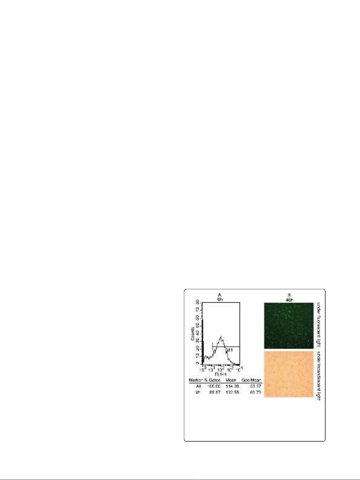
fluorescence with 100 nM fluorescent siRNA (Figure 1).
Figure 1 Efficient delivery of siRNA into lung adenocarcinoma cells.
(A): Detection of transfection efficiency by flow cytometry. Transfection
efficiency was maintained at over 85% for 6 h post-transfection. (B):
Detection of transfection efficiency by fluorescence microscopy. High
efficiency of transfection with fluorescent siRNA (green) in A549 cells
were easily identified for 48 h post-transfection (×100).
Xu et al.Journal of Experimental & Clinical Cancer Research 2011, 30:63
http://www.jeccr.com/content/30/1/63
Page 3 of 11
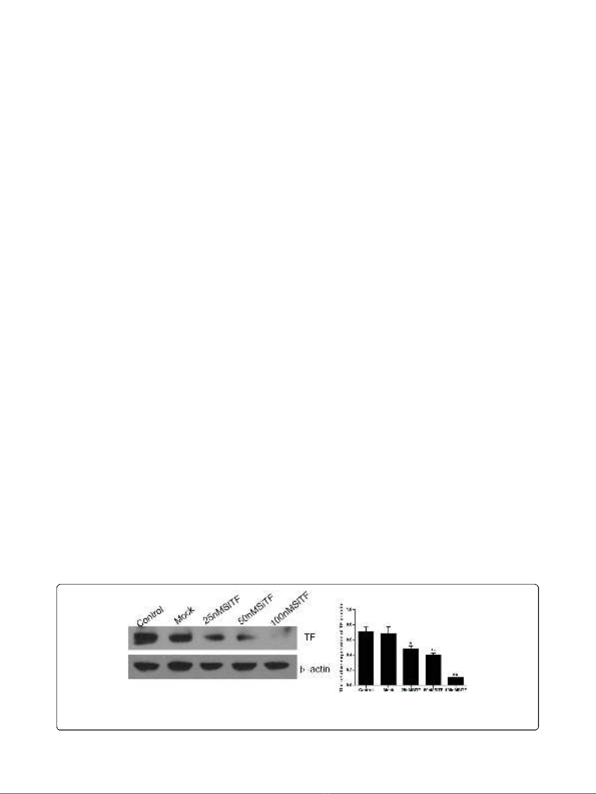
When cells were treated with TF-targeting siRNA
(25 nM, 50 nM and 100 nM SiTF) and the scramble
siRNA (Mock, 100 nM) for 48 h, the mRNA and protein
expressions of TF were examined by RT- PCR and Wes-
tern blot. As shown in Figure 2 and Figure 3, the Mock
did not affect the expression levels of TF, but in 25 nM,
50 nM and 100 nM SiTF groups, compared with mock,
the TF expression decreased at both protein and mRNA
levels. Specially, 100 nM SiTF indicated a 80-85% reduc-
tion of TF expression in A549 cells. These results
demonstrated that the TF-targeting siRNA was efficient
to knock down the expression of TF in A549 cells.
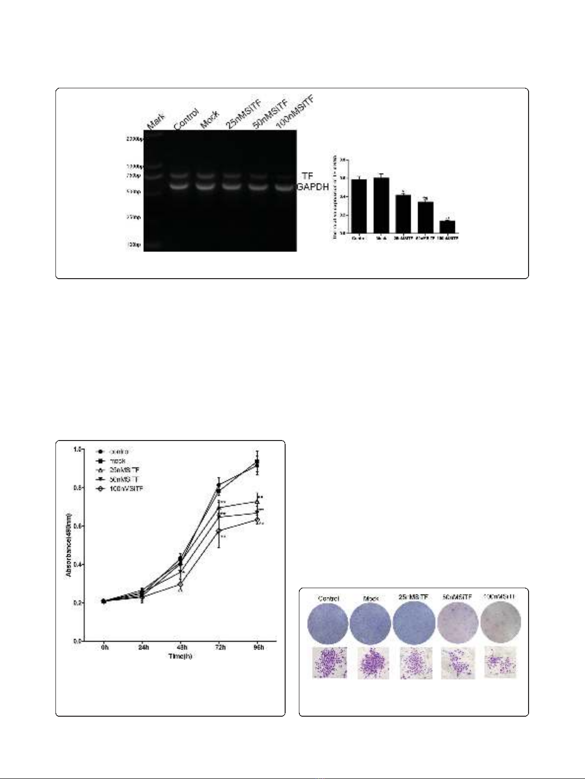
Inhibition of cell proliferation and colony formation by
TF-siRNA
Since previous studies have shown that the expression of
TF associated with tumor growth [20-22], the effect of
TF siRNA on lung adenocarcinoma cell proliferation
was determined by MTT assay. As shown in Figure 4,
after 24 h-96 h transfection of TF siRNA into A549
cells, cell proliferation was remarkably inhibited in a
time- and dose-dependent manner, when compared
with control and mock groups. Inhibition of cell prolif-
eration at 50 nM and100 nM began at 48 h post-trans-
fection,butat25nMwasobservedat72hpost-
transfection, and higher concentrations of TF siRNA
had greater effects. In addition, the colony formation
assay further revealed effects of TF knockdown on
growth properties of A549 cells. 50 nM and100 nM
SiTF groups, but not 25 nM SiTF group had lower posi-
tive colony formation than control and mock groups,
and it also seemed to depend on doses (Figure 5 and
Figure 6). Overall, down-regulation of TF by siRNA
resulted in a negative effect on growth of lung adenocar-
cinoma cells.
Attenuation of the migration/invasion ability by TF-siRNA
Tumor cell migration and invasion are two critical steps
in cancer metastatic process [23]. To verify the effect of
TF-siRNA on the migration ability, A549 cells were
tested by wound healing assay and the mobility assay.
Figure 7 and Figure 8 show that the cells in 50 nM and
100 nM SiTF groups demonstrated an attenuated capa-
city of impaired migration, when compared to control
and mock groups. Moreover, untreated and transfected
cells were seeded on transwell chambers with uncoated
filters. After incubation for 24 h, the motility potential
of transfected cells at 50 nM and 100 nM TF-siRNA
was significantly suppressed (Figure 9 and Figure 10). In
addition, the invasion assay using Matrigel-coated
Transwell chambers showed that 50 nM and 100 nM
TF-siRNA transfected cells that passed through the
Matrigel-coated membranes were much more than par-
ental cells and the cells transfected with scrambled
siRNA, and it indicated that the invasive capacity was
markedly decreased (Figure 11 and Figure 12). These
results suggested that TF-siRNA attenuated the meta-
static potential of lung adenocarcinoma cells in vitro.
Promoted apoptosis in A549 cells by TF-siRNA
To evaluate further whether knockdown of TF induces
A549 cells apoptosis, at 48 h after transfection, the cells
were harvested and analyzed by flow cytometry. As
shown in Figure 13, the apoptosis rates of 25 nM, 50
nM and 100 nM SiTF groups were 7.0%, 9.0% and
16.0%, respectively, which were higher than 4.0% in con-
trol and 4.8% in mock groups, and indicated a dose-
dependent increase.
Molecular mechanisms of the antitumor effects by
TF-siRNA
The protein from transfected cells was extracted to
examine the effects of TF-siRNA on some important
cytokines and signaling molecules. After 48 h of trans-
fection, the protein relative expression levels of phos-
phorylated Erk1/2 and PI3K in 100 nM SiTF group and
phosphorylated Akt in 25 nM, 50 nM and 100 nM SiTF
groups were decreased, while that in control and mock
groups had no differences (Figure 14 and Figure 15).
Furthermore, compared to control and mock groups,
Figure 2 TF-siRNA suppressed the TF protein expression in lung adenocarcinoma cells. 48 h after transfection, the concentration of 100
nM TF-siRNA (100 nM SiTF group) was identified as the most efficient to knock down the expression of TF by Western blot. *P< 0.05, **P< 0.01
versus mock.
Xu et al.Journal of Experimental & Clinical Cancer Research 2011, 30:63
http://www.jeccr.com/content/30/1/63
Page 4 of 11
transfection with high concentrations of 50 nM and 100
nM TF-siRNA suppressed the MMP-9/-2 expression
(Figure 16), and the protein expression of VEGF of 100
nM SiTF group was decreased (Figure 17). These data
demonstrated that knockdown of TF by siRNA may
inhibit Erk1/2 MAPK, PI3K/Akt signaling pathway,
MMP-9/-2 and VEGF, which all play an important role
in tumor progress.
Inhibition of tumor growth of lung adenocarcinoma cells
in nude mice by TF-siRNA
Intratumoral injection with TF-siRNA was performed to
investigate whether TF-siRNA had the effect of inhibi-
tion on tumor growth in vivo. A nude-mouse model of
human lung adenocarcinoma xenograft was established,
and when the tumor volume reached 50-100 mm
3
,
intratumoral treatment with TF-siRNAs was started and
repeated every 5 days for a total of 5 times. As shown in
Figure 18A, the tumor volume of SiTF group from days
22 to the end was significantly smaller than control and
mock groups, whereas there was no statistical difference
between control group and mock group during the
experiment. All mice were sacrificed on the 42nd day,
and the final tumor volume and weight in SiTF group
(209.6 ± 97.6 mm
3
and 0.21 ± 0.10 g, n = 5) were mark-
edly smaller than that in control group (600.8 ± 182.0
mm
3
and 0.59 ± 0.18 g, n = 5) and mock group (513.8
± 112.6 mm
3
and 0.52 ± 0.12 g, n = 5) (Figure 18 and
Figure 19). In addition, the relative protein expression of
TF in SiTF group was decreased significantly, but there
was no statistical significance between control group
and mock group (Figure 20). After all, these results
Figure 4 Knockdown of TF with TF-siRNA inhibited cell
proliferation of lung adenocarcinoma cells in vitro. TF-siRNAs
transfected A549 cell growth was significantly attenuated in a time-
and dose-dependent manner compared with mock. *P< 0.05, **P<
0.01 versus mock.
Figure 3 TF-siRNA suppressed the mRNA expression in lung adenocarcinoma cells. The concentration of 100 nM TF-siRNA (100 nM SiTF
group) was identified as the most efficient to knock down the expression of TF by RT-PCR. *P< 0.05, **P< 0.01 versus mock.
Figure 5 Knockdown of TF with TF-siRNA inhibited colony
formation of lung adenocarcinoma cells in vitro. Representative
images of the colony formation assay were shown.
Xu et al.Journal of Experimental & Clinical Cancer Research 2011, 30:63
http://www.jeccr.com/content/30/1/63
Page 5 of 11


