RESEARC H Open Access
Molecular characterization of genome segments
1 and 3 encoding two capsid proteins of
Antheraea mylitta cytoplasmic polyhedrosis virus
Mrinmay Chakrabarti, Suvankar Ghorai, Saravana KK Mani, Ananta K Ghosh
*
Abstract
Background: Antheraea mylitta cytoplasmic polyhedrosis virus (AmCPV), a cypovirus of Reoviridae family, infects
Indian non-mulberry silkworm, Antheraea mylitta, and contains 11 segmented double stranded RNA (S1-S11) in its
genome. Some of its genome segments (S2 and S6-S11) have been previously characterized but genome
segments encoding viral capsid have not been characterized.
Results: In this study genome segments 1 (S1) and 3 (S3) of AmCPV were converted to cDNA, cloned and
sequenced. S1 consisted of 3852 nucleotides, with one long ORF of 3735 nucleotides and could encode a protein
of 1245 amino acids with molecular mass of ~141 kDa. Similarly, S3 consisted of 3784 nucleotides having a long
ORF of 3630 nucleotides and could encode a protein of 1210 amino acids with molecular mass of ~137 kDa.
BLAST analysis showed 20-22% homology of S1 and S3 sequence with spike and capsid proteins, respectively, of
other closely related cypoviruses like Bombyx mori CPV (BmCPV), Lymantria dispar CPV (LdCPV), and Dendrolimus
punctatus CPV (DpCPV). The ORFs of S1 and S3 were expressed as 141 kDa and 137 kDa insoluble His-tagged
fusion proteins, respectively, in Escherichia coli M15 cells via pQE-30 vector, purified through Ni-NTA
chromatography and polyclonal antibodies were raised. Immunoblot analysis of purified polyhedra, virion particles
and virus infected mid-gut cells with the raised anti-p137 and anti-p141 antibodies showed specific
immunoreactive bands and suggest that S1 and S3 may code for viral structural proteins. Expression of S1 and S3
ORFs in insect cells via baculovirus recombinants showed to produce viral like particles (VLPs) by transmission
electron microscopy. Immunogold staining showed that S3 encoded proteins self assembled to form viral outer
capsid and VLPs maintained their stability at different pH in presence of S1 encoded protein.
Conclusion: Our results of cloning, sequencing and functional analysis of AmCPV S1 and S3 indicate that S3
encoded viral structural proteins can self assemble to form viral outer capsid and S1 encoded protein remains
associated with it as inner capsid to maintain the stability. Further studies will help to understand the molecular
mechanism of capsid formation during cypovirus replication.
Background
Cytoplasmic polyhedrosis virus or CPV of the genus
Cypovirus of Reoviridae family [1,2] infects the midgut
of the wide range of insects belonging to the order
Diptera, Hymenoptera and Lepidoptera [3,4]. Like
other members of Reoviridae,CPVgenomeisalso
composed of 10 double stranded RNA segments
(dsRNA) (S1-S10) [2]. A small eleventh segment (S11)
has been reported in some cases such as Bombyx mori
CPV (BmCPV) [5] and Trychoplusia ni CPV (TnCPV)
[6]. Each dsRNA segment is composed of a plus
mRNA strand and it’s complementary minus strand in
an end to end base pair configuration except for a pro-
truding 5′cap on the plus strand. On the basis of elec-
trophoretic migration patterns of the dsRNA segments
in agarose or acrylamide gels, CPVs have been classi-
fied into 16 different types [1,7]. CPVs are self compe-
tent for transcription, possessing all the enzymes
necessary for mRNA synthesis and processing [8].
BmCPV, the type Cypovirus, has a single layer capsid
made up of 120 copies of the major capsid protein,
* Correspondence: aghosh@hijli.iitkgp.ernet.in
Department of Biotechnology, Indian Institute of Technology Kharagpur,
Kharagpur 721302, West Bengal, India
Chakrabarti et al.Virology Journal 2010, 7:181
http://www.virologyj.com/content/7/1/181
© 2010 Chakrabarti et al; licensee BioMed Central Ltd. This is an Open Access article distributed under the terms of the Creative
Commons Attribution License (http://creativecommons.org/licenses/by/2.0), which permits unrestricted use, distribution, and
reproduction in any medium, provided the original work is properly cited.

VP1, which is decorated with 12 turrets on its icosahe-
dral vertices [9,10]. These hollow turrets are involved
in post-transcriptional processing of viral mRNA and
provide a channel through which newly synthesized 5′
capped viral RNA are released from the capsid into the
cytoplasm of infected cells [10,11]. After translation of
this mRNA into capsid, polymerase and other proteins,
they assembled into viral procapsid within which one
copy of each genome segments plus polarity RNA are
packaged and replicated to form dsRNA. CPV capsids
thus formed can be released as non-occluded virus
particles to directly infect fresh neighboring cells or
occluded in a viral protein matrix called polyhedrin to
form polyhedra [12]. It has been reported that VP1
protein, encoded by genome segment 1 of BmCPV,
can self assemble to form single shelled virus like par-
ticles (VLPs) [13,14] and their stability is maintained
by interaction with VP3 and VP4 proteins encoded by
genome segments 3 and 4, respectively [15,16]. Recent
cryo-electron microscopic study has shown the region
of capsid protein directly interacting with viral RNA
indicating the role of capsid in RNA packaging, repli-
cation and transcription [17]. Therefore, understanding
the assembly of capsid not only provides insight into
in the virus life cycle but also helps to develop
mechanism for the disruption of virus assembly for
therapeutic application [18]. But besides BmCPV, cap-
sids of other CPVs have not been studied well
although all the genome segments of DpCPV, LdCPV
and TnCPV have been cloned and sequenced [6,19-21].
Antheraea mylitta cytoplasmic polyhedrosis virus
(AmCPV) is one of the most widespread pathogens of
Indian non-mulberry silkworm, A. mylitta. CPV-infected
A. myllita larvae develop chronic diarrhea that even-
tually leads to a condition known as “Grasserie”and
ultimate death [22]. Almost 20-30% larval mortality
occurs annually due to this virus attack [22]. We have
previously characterized the structure of AmCPV by
electron microscopy and its genome by electrophoresis
which reveals that it is similar to that of a type- 4 CPV
and consists of 11 ds RNA molecules [23]. We have also
reported that the genome segments 6, 7, 8 of AmCPV
encode viral structural proteins [24-26], segment 2
encodes viral RNA dependent RNA polymerase [27],
segment 9 encodes a nonstructural protein, NSP38, hav-
ing RNA binding property [28], segment 10 codes for
polyhedrin [29] and segment 11 does not code for any
protein [26]. But the genome segments encoding viral
capsid proteins have not been characterized. Here, we
report molecular cloning, sequencing and expression of
S1 and S3 of AmCPV in E. Coli via bacterial expression
vector as well as in insect cells using a baculovirus sys-
tem and show by functional analysis that S3 encoded
protein can self assemble into capsid and S1 encoded
protein remains associated with the capsid to maintain
its stability.
Results and discussion
Genetic analysis of AmCPV S1 and S3
AmCPV S1 and S3 RNA were isolated, converted to
cDNA and cloned into pCR-XL-TOPO and the total
nucleotide sequences were determined in both forward
and reverse directions. S1 consisted of 3852 nucleotides
with one long ORF of 3735 nucleotides and could
encode a protein of 1245 amino acids with molecular
mass of ~141 kDa (p141). Thirty four nucleotides
upstream of translation initiation codon (ATG) and 80
nucleotides downstream of translation stop codon
(TGA) were present at untranslated sequences (Gen-
bank accession No: HM230690). Similarly, S3 consisted
of 3784 nucleotides having a long ORF of 3630 nucleo-
tides and could encode a protein of 1210 amino acids
with molecular mass of ~137 kDa (p137). Twenty seven
nucleotides upstream of translation initiation codon
(ATG) and 124 nucleotides downstream of translation
stop codon (TGA) were present as untranslated
sequences (Genbank accession No: HM230691). Cloning
of S1 and S3 was confirmed by northern analysis of
total AmCPV RNA using cloned S1 and S3 cDNA as
probes (data not shown).
BLASTP results showed 22%, 23% and 27% homology
of AmCPVS1 encoded p141 with segment 3 encoded
proteins VP3, VP2 and a hypothetical protein of
BmCPV1, DpCPV1 and LdCPV14, respectively
[13,20,21]. Function of VP3 protein of BmCPV1 is not
exactly known but probably codes for spike protein [13].
Therefore it is suggested that AmCPV S1 may also code
for a minor capsid protein which is probably involved in
spike formation. AmCPV S1 contained seventeen N-
linked glycosylation sites, two cAMP- and cGMP-depen-
dent protein kinase phosphorylation sites, twenty casein
kinase II phosphorylation sites, twelve N-myristoylation
sites, fourteen protein kinase C phosphorylation sites
and two tyrosine kinase phosphorylation sites. Second-
ary structure prediction with PHD and GOR4 showed
that 36.54% of the residues are likely to form a-helices,
25.69% would form extended sheets and 37.77% would
form random coils. But their functional significance
remains to be determined.
BLASTP results also showed 20-23% homology of
AmCPV S3 encoded p137 with segment 1 encoded
major capsid protein, VP1, of BmCPV, DpCPV and
LdCPV indicating that AmCPVS3 may code for major
capsid protein of AmCPV. AmCPV S3 contained eight
N-linked glycosylation sites, one cAMP- and cGMP-
dependent protein kinase phosphorylation site, 14 pro-
tein kinase C phosphorylation sites, 19 casein kinase II
phosphorylation sites, 13 N-myristoylation sites and
Chakrabarti et al.Virology Journal 2010, 7:181
http://www.virologyj.com/content/7/1/181
Page 2 of 11
one prokaryotic membrane lipoprotein lipid attach-
ment site. Secondary structure prediction with PHD
and GOR4 showed that 28% of the residues are likely
to form a-helices, 14.9% would form extended sheets
and 57.1% would form random coils. But the correla-
tion between this structure and function remains to be
made. In both the genes the 5′terminal sequence
AGTAAT and 3’terminal sequence AGAGC were
found to be the same as the 5′and 3’terminal
sequences found in AmCPV genome segments 2, 6, 7,
8 and 10 indicating that the genome structure of this
CPV may follow the same pattern as found in other
CPVs [6,19-21,30].
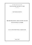
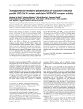
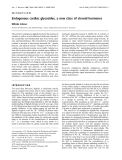
Phylogenetic analysis of AmCPV S1 and S3 amino acid
sequences with other viruses in the Reoviridae showed its
close relatedness with some members of the cypovirus
such as BmCPV-1, DpCPV and LdCPV (Fig. 1A &1B)
and indicates that all these cypoviruses mayhavebeen
originated from a common ancestral insect virus.
Figure 1 Phylogenetic analysis of AmCPV S1 (A) and AmCPV S3 (B) encoded proteins with other members of the Reoviridae.The
number at each node represents bootstrap value of 100 replicates. GenBank accession numbers are shown in parenthesis.
Chakrabarti et al.Virology Journal 2010, 7:181
http://www.virologyj.com/content/7/1/181
Page 3 of 11
Analysis of recombinant AmCPV S1 and S3 encoded
proteins expressed in E. coli and insect cells
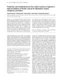
AmCPV S1 and S3 were expressed in E. coli M15 cells
as insoluble 141 kDa (Fig. 2A, lanes 3 & 4) and 137 kDa
(Fig. 2B, lanes 2 & 3) proteins, respectively. Polyclonal
antibodies were raised in a rabbit against purified p141
and p137, and titered as 10
-4
by ELISA.
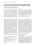
Sf9 cells infected with S1 and S3 recombinant bacu-
lovirus expressed these proteins in soluble form as 141
and 137 kDa, respectively [Fig 3A and 4A (lane 1)].
This was confirmed by immunoblot analysis (Fig. 3B
and 4B, lane 1). Expression of predicted same size pro-
teins both in bacteria and insect cells indicate that
although a number of glycosylation sites are present in
both these genes but they are not used for post trans-
lational modification. In E. coli M15 cells the expressed
proteins might not fold properly into correct confor-
mation and thus the incorrectly folded protein may
have aggregated to produce insoluble inclusion bodies
but in insect (Sf9) cells via baculovirus expression sys-
tem due to proper folding soluble proteins are
produced.
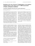
To determine function of AmCPV S1 and S3 encoded
proteins, immunoblot analysis was done with the midgut
of uninfected and virus-infected larvae, polyhedra and
virion particles using purified polyclonal anti-p141 and
anti-p137 antibodies. Major immunoreactive bands of
141 kDa and 137 kDa (Fig. 3 &4, lanes 3, 4 and 5) were
observed in infected midgut, purified polyhedra as well
as virus particles, but not in uninfected midgut (lane 2)
indicating that they might code for two viral structural
proteins.
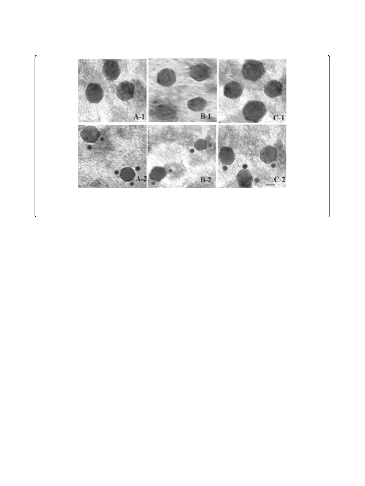
Transmission Electron Microscopic (TEM) analysis of virus
like particles
To visualize the formation of virus like particles (VLPs)
in recombinant baculovirus infected Sf9 cells and to
confirm the identity of their protein content, VLPs were
purified from infected cells and immunogold staining of
the particles were performed using rabbit anti-p141 or
anti-p137 antibodies. As shown by TEM analysis (Fig. 5.
A-2, B-2, C-2), native AmCPV, recombinant VLP from
Sf 9 cells infected with AmCPV S3 baculovirus recombi-
nants alone or, Sf9 cells co-infected with AmCPV S1
and S3 baculovirus recombinants were specifically
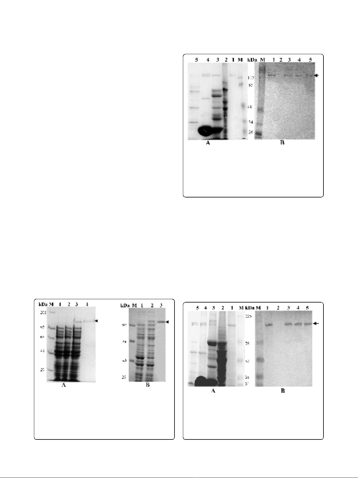
Figure 2 (A) Analysis of E. coli M15 expressed AmCPV S1
encoded protein by SDS-8% PAGE. Lane M, Molecular weight
marker (Bangalore Genei); lane 1, uninduced cell lysate; lane 2,
induced cell supernatant; lane 3, induced cell pellet; lane 4, Ni-NTA
purified protein. (B) Analysis of E. coli M15 expressed AmCPV S3
encoded protein by SDS-8% PAGE. Lane M, Molecular weight
marker (Bangalore genei); lane 1, uninduced cell lysate; lane 2,
induced cell lyaste; lane 3, Ni-NTA purified protein.
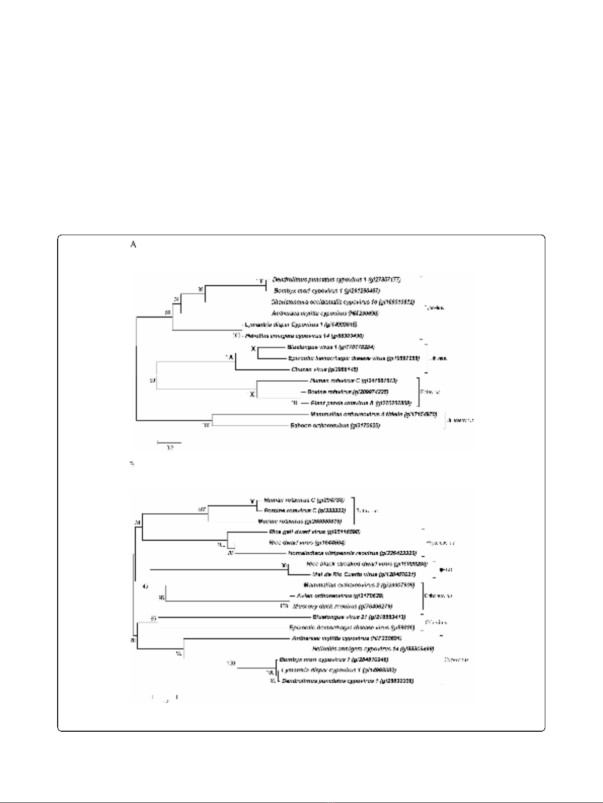
Figure 3 Immunoblot analysis of AmCPV S1 encoded proteins
using anti-p141 polyclonal antibody. (A) SDS-8% PAGE and (B)
Western Blot. Lane M, Prestained protein molecular weight marker
(Fermentas); lane 1, purified insect cell expressed recombinant p141
protein; lane 2, uninfected midgut of A. mylitta; lane 3, infected
midgut of A. mylitta; lane 4, purified polyhedra and lane 5, purified
virion particle. Arrow indicates the position of immunoreactive
protein.
Figure 4 Immunoblot analysis of AmCPV S3 encoded proteins
using anti-p137 polyclonal antibody. (A) SDS-8% PAGE and (B)
Western Blot. Lane M, Prestained molecular weight marker (GE); lane
1, purified insect cell expressed recombinant p137 protein; lane 2,
uninfected midgut of A. mylitta; lane 3, infected midgut of A.
mylitta; lane 4, purified polyhedra; and lane 5, purified virion particle.
Arrow indicates the position of immunoreactive protein
Chakrabarti et al.Virology Journal 2010, 7:181
http://www.virologyj.com/content/7/1/181
Page 4 of 11
labeled with rabbit anti-p137 antibody conjugated gold
particles. No gold particle labeling was observed when
anti-p141 antibody was used (data not shown). Also no
VLP formation was observed in cells infected with
AmCPVS1 recombinant baculovirus alone. These results
indicate that AmCPV S3 encoded protein alone has the
ability to self assemble for the formation of single
shelled particle (capsid) without the assistance of any
other structural protein of AmCPV. Similar capsid for-
mation has been reported for BmCPV S1 encoded VP1
protein [14]. No gold particle labeling in VLPs produced
from Sf9 cells co-infected with AmCPV S1 and S3
recombinants using anti-p141 antibody may be due to
presence of S1 encoded protein in the inner side of cap-
sid where antibody can not access or absence of epitope
(exposed outside) specific antibody in the raised polyclo-
nal antibody.
Immunoblot analysis of VLPs
To further confirm the protein content of these VLPs
obtained from recombinant baculovirus infected Sf9
cells, immunoblot analysis was performed using anti-
p137 and anti-p141 antibodies. Immunoblot study using
anti-p137 antibody (Fig. 6B) showed a single major
immunoreactive band at 137 kDa in purified VLPs from
cells infected with AmCPV S3 baculovirus recombinants
(lane 1), purified VLPs from cells co-infected with both
AmCPV S1 and S3 baculovirus recombinants (lane 2),
purified p137 protein (lane 3) and native virion particles
(lane 5). Similar immunoblot study, using anti-p141
antibody showed a single major immunoreactive band at
141 kDa in purified VLPs obtained from cells expressing
both AmCPV S1 and S3 (Fig. 6C, lane 2), purified
recombinant p141 protein (lane 4) and native virion par-
ticles (lane 5). Since in SDS-PAGE, after Coomassie blue
staining two bands (137 kDa and 141 kDa) were
observed in purified VLPs from cells co-infected with
both AmCPV S1 and S3 baculovirus recombinants (lane
2), and also in purified native virion particles (lane 5)
and reacted with both anti-p141 and p137 antibodies,
these results indicate that p137 is involved in the forma-
tion capsid outer shell and p141 is associated in the
inner side of capsid (VLPs). Three dimensional structure
of BmCPV has shown presence of spike molecules and
transcription enzyme complexes along the icosahedral
five fold axis both inside and outside of the core parti-
cles [10,16]. Similar studies are required to understand
the association of AmCPV S1 and S3 encoded proteins
in the viral capsid.
Comparison of stability of native virion and virus like
particles at different pH
Transmission electron microscopic studies of native vir-
ions and recombinant VLPs at different pH showed that
VLPs are more stable in alkaline condition rather than
acidic pH (Fig. 7). Most of VLPs maintained their intact
structure at pH-12 whereas totally disintegrated at
below pH-4. At any given pH native virion particles
were found to be more stable than VLPs made up of
p137 or p137 and p141 together (Table 1). But VLPs
Figure 5 Electron micrographs of uranyl acetate-stained native and recombinant VLPs of AmCPV. (A) Native AmCPV particles; (B)
Recombinant VLPs expressing AmCPV S3 encoded protein; (C) Recombinant VLPs expressing AmCPV S1 and AmCPV S3 encoded proteins. Upper
panel (A-1, B-1 and C-1) shows the purified particles in 20 mM PBS, pH 7.3 and lower panel (A-2, B-2, and C-2) shows immunogold staining of
these particles. Bar, 20 nm.
Chakrabarti et al.Virology Journal 2010, 7:181
http://www.virologyj.com/content/7/1/181
Page 5 of 11