


Chemical constituents of the leaves of Taxus wallichiana (Taxaceae)
lượt xem 4
download
 Download
Vui lòng tải xuống để xem tài liệu đầy đủ
Download
Vui lòng tải xuống để xem tài liệu đầy đủ
From CHCl3-soluble extract of the leaves of Taxus wallichiana (Taxaceae), collected in Lam Dong Province, five compounds were isolated and identified as sciadopitysin, ginkgetin, (S)-(+)-rhododenol, (+)-erythro-N-benzoyl3-phenylisoserine methyl ester, and (−)-α-conidendrin (5). Their chemical structures were determined by 1D and 2D NMR spectra and comparison with published data. Except for (−)-α-conidendrin, these remaining compounds were first reported of Taxus wallichiana. Among these compounds, ginkgetin and (−)-αconidendrin showed significant α-glucosidase inhibitory activity with the IC50 values of 98.5, and 172.1 μM, respectively.
Bình luận(0) Đăng nhập để gửi bình luận!
Nội dung Text: Chemical constituents of the leaves of Taxus wallichiana (Taxaceae)
- Received: 18 February 2023 Revised: 30 March 2023 Accepted: 24 July 2023 DOI: 10.1002/vjch.202300059 RESEARCH ARTICLE Chemical constituents of the leaves of Taxus wallichiana (Taxaceae) Thinh Duc Le1,2 Da Vy Thi Le1,2 Tu Hoai Tran1,2 Phu Hoang Dang1,2 Tho Huu Le1,2 Mai Thanh Thi Nguyen1,2 Nhan Trung Nguyen1,2 1 Faculty of Chemistry, University of Science, Ho Chi Minh City, Vietnam Abstract 2 Management Agency, Vietnam National From CHCl3 -soluble extract of the leaves of Taxus wallichiana (Taxaceae), col- University Ho Chi Minh City, Ho Chi Minh City, lected in Lam Dong Province, five compounds were isolated and identified as Vietnam sciadopitysin (1), ginkgetin (2), (S)-(+)-rhododenol (3), (+)-erythro-N-benzoyl- 3-phenylisoserine methyl ester (4), and (−)-α-conidendrin (5). Their chemical Correspondence Nhan Trung Nguyen, Faculty of Chemistry, structures were determined by 1D and 2D NMR spectra and comparison with pub- VNUHCM-University of Science, 227 Nguyen Van lished data. Except for (−)-α-conidendrin, these remaining compounds were first Cu Street, Ward 4, District 5, Ho Chi Minh City reported of Taxus wallichiana. Among these compounds, ginkgetin (2), and (−)-α- 72711, Vietnam. Email: ntnhan@hcmus.edu.vn conidendrin (5) showed significant α-glucosidase inhibitory activity with the IC50 values of 98.5, and 172.1 μM, respectively. Funding information Vietnam National University, Ho Chi Minh City KEYWORDS (VNU-HCM), Grant/Award Number: α-glucosidase inhibitory activity, Taxaceae, Taxus wallichiana 562-2022-18-04 1 INTRODUCTION showed strong α-glucosidase inhibitory activity, with the IC50 value of 0.30 μg/mL. Hence, an additional fractionation Taxus wallichiana Zucc., commonly known as the Wallich’s study was undertaken to examine its chemical constituents, yew, is an evergreen coniferous tree of medium size, reach- resulting in the isolation of five compounds. This work ing heights between 10 to 20 m. It is native to several coun- describes isolating and elucidating the chemical structures tries in Asia, including Afghanistan, India, Pakistan, Nepal, of these compounds by spectroscopic methods and their China, Bhutan, Indonesia, Malaysia, Myanmar, Vietnam, and α-glucosidase inhibitory activity evaluation. the Philippines.1 In Vietnam, the wood powder derived As part of an ongoing investigation into the screening from Taxus wallichiana is utilized in traditional medicine of medicinal plants for their potential α-glucosidase for various purposes, such as treating high fever, diabetes, inhibitory activity,4,5 the CHCl3 -soluble extract from and indigestion, providing anti-inflammatory effects, and T. wallichiana leaves was found to exhibit a remarkably alleviating pain. Numerous previous studies have high- strong α-glucosidase inhibitory activity, with an IC50 value lighted the medicinal properties of T. wallichiana, includ- of 0.30 μg/mL. This promising finding prompted a subse- ing its antipyretic, antibacterial, antifungal, analgesic, and quent fractionation study focused on isolating the chemical anticonvulsant characteristics.2 These beneficial proper- constituents responsible for this activity, which resulted in ties are attributed to the tree’s abundant chemical con- the identification of five compounds. stituents, such as taxane diterpenoids,3 flavonoids,2 and This research involved the isolation and elucidation of lignans.2 the chemical structures of these five compounds using In a continued study on the screening of medicinal plants spectroscopic methods. Furthermore, their α-glucosidase for α-glucosidase inhibitory activity,4,5 it was found that inhibitory activity was evaluated to determine their poten- the CHCl3 -soluble extract of the leaves of T. wallichiana tial effectiveness in this regard. © 2024 Vietnam Academy of Science and Technology and Wiley-VCH GmbH. Vietnam J. Chem. 2024;62:37–42. wileyonlinelibrary.com/journal/vjch 37
- 25728288, 2024, 1, Downloaded from https://onlinelibrary.wiley.com/doi/10.1002/vjch.202300059 by Readcube (Labtiva Inc.), Wiley Online Library on [01/05/2024]. See the Terms and Conditions (https://onlinelibrary.wiley.com/terms-and-conditions) on Wiley Online Library for rules of use; OA articles are governed by the applicable Creative Commons License 38 LE ET AL. 2 MATERIALS AND METHODS (10 → 100% MeOH) and followed by preparative TLC with CHCl3 –acetone (9:1), to afford compound 1 (4.0 mg). Sub- 2.1 General procedures fraction Fr.D4.4 (609 mg) was separated by silica gel column chromatography eluted with n-hexane–acetone (0 → 100% The NMR (Nuclear Magnetic Resonance) spectra were acetone) and followed by preparative TLC with toluene- acquired using a Bruker Avance spectrometer, operating at EtOAc-AcOH (88:10:2), to obtain compounds 2 (3.0 mg) and 600 MHz for the 1 H NMR spectrum and 150 MHz for the 3 (3.5 mg). 13 C NMR spectrum. Deuterated solvents were employed Fraction Fr.E was chromatographed with n-hexane– as internal standards, and the chemical shifts were repre- acetone (0 → 100% acetone) and MeOH (0 → 100% sented in δ values. Column chromatography was performed MeOH) mixtures, to yield five subfractions (Fr.E1–E5). Sub- using silica gel 60 F254 (particle size 0.040–0.063 mm, Merck fraction E3 (6.9 g) was continued to be subjected to normal Kieselgel), silica gel RP-18 (Merck), and resin chromatogra- phase chromatography with n-hexane–EtOAc (0 → 100% phy utilizing Diaion HP20. For thin-layer chromatography EtOAc) and MeOH (0 → 100% MeOH) mixtures, to yield (TLC), precoated plates of Kieselgel 60 F254 or RP18 with a 5 subfractions (Fr.E3.1–E3.5). Subfraction Fr.E3.3 (3 g) was thickness of 0.25 mm (Merck) were used. purified with n-hexane–EtOAc (0 → 100% EtOAc) and MeOH (0 → 100% MeOH) mixtures, to yield 4 subfrac- tions (Fr.E3.3.1–E3.3.4). Subfraction Fr.E3.3.2 (73.5 mg) is 2.2 Plant material passed silica gel column chromatography, eluted with n-hexane–CHCl3 –isopropanol (60:30:1), to obtain com- In January 2021, the leaves of Taxus wallichiana Zucc. were pounds 4 (4.0 mg) and 5 (3.0 mg). gathered in the Don Duong District, Lam Dong Province, Fraction F (11.62 g) was chromatographed with n- Vietnam. Anh Tuan Dang-Le from the Faculty of Biology and hexane–EtOAc (0 → 100% EtOAc) and EtOAc–MeOH Biotechnology at the University of Science, Ho Chi Minh (0 → 100% MeOH) mixtures, to yield 4 subfractions City, Vietnam, conducted the identification of the scientific (Fr.F1–F4). Subfraction F4 (1.4 g) was purified with name. A voucher specimen (MLTD0005) has been appropri- n-hexane–acetone (0 → 100% acetone) and MeOH ately stored and preserved at the Department of Organic (0 → 100% MeOH) mixtures to afford compound 5 (5.0 mg) Chemistry, Faculty of Chemistry, VNUHCM−University of (Figure 1). Science. Sciadopitysin (1): HRESIMS: m/z 603.1269 [M+Na]+ (C33 H24 O10 Na+ , 603.1267). 1 H NMR (600 MHz, pyridine-d5 ): δH (J in Hz) 3.68 (3H, s, 4‴-OCH3 ), 3.78 (3H, s, 4′-OCH3 ), 3.88 2.3 Extraction and isolation of (3H, s, 7-OCH3 ), 6.62 (1H, d, 2.4, H-6), 6.74 (1H, d, 2.4, H-8), compounds 7.03 (1H, s, H-6″), 7.04 (1H, s, H-3), 7.06 (2H, d, 8.5, H-3‴, H-5‴), 7.17 (1H, s, H-3″), 7.46 (1H, d, 8.8, H-5′), 7.80 (2H, d, Seven kilograms of powdered dried leaves from Taxus 8.5, H-2‴, H-6‴), 8.24 (1H, dd, 8.8, 2.5, H-6′), 8.53 (1H, d, 2.5, wallichiana Zucc. were subjected to extraction with 1.5 L H-2′). 13 C NMR (150 MHz, pyridine-d5 ): δC 183.0 (C-4), 182.9 of MeOH, using a ratio of 200 g of leaves to 1.5 L of (C-4″), 165.8 (C-7), 164.3 (C-2, C-2″), 163.8 (C-7″), 162,8 MeOH. The extraction was carried out through reflux for (C-4′, C-5″, 4‴), 161.5 (C-5), 158.1 (C-9″), 156.0 (C-9), 131.9 3 h, repeated four times, producing 1 kg of methanolic (C-2′), 128.6 (C-2‴, C-6‴), 128.5 (C-6′), 123.8 (C-1‴), 123.5 extract. This methanolic extract was then suspended with (C-1′), 123.3 (C-3′), 114.9 (C-3‴, C-5‴), 111.9 (C-5′), 105.8 H2 O and sequentially partitioned using n-hexane, CHCl3 , (C-10″), 104.9 (C-10), 104.7 (C-3″, C-8″), 104.1 (C-3), 99.6 and EtOAc. This partitioning process led to the isolation of (C-6″), 98.6 (C-6), 92.9 (C-8), 56.0 (7-OCH3 ), 55.9 (4′-OCH3 ), the n-hexane-soluble fraction (110 g), chloroform-soluble 55.4 (4‴-OCH3 ). fraction (200 g), and EtOAc-soluble fraction (98 g). The Ginkgetin (2): HRESIMS: m/z 589.1115 [M+Na]+ CHCl3 -soluble fraction was subjected to a silica gel column, (C32 H22 O10 Na+ , 589.1111). 1 H NMR (600 MHz, pyridine-d5 ): eluted with n-hexane–EtOAc (0 → 100% EtOAc) and EtOAc– δH (J in Hz) 3.73 (3H, s, 7-OCH3 ), 3.78 (3H, s, 4′-OCH3 ), 6.56 MeOH (0 → 100% MeOH) mixtures, to yield 10 fractions (1H, d, 1.8, H-6), 6.68 (1H, d, 1.8, H-8), 6.93 (1H, s, H-3″), (Fr.A–Fr.J). 6.99 (1H, s, H-6″), 7.07 (1H, s, H-3), 7.17 (2H, d, 9.0, H-3‴, Fraction Fr.D (7.49 g) was chromatographed over a silica H-5‴), 7.33 (1H, d, 8.4, H-5′), 7.74 (2H, d, 9.0, H-2‴, H-6‴), gel column, with elution using a gradient of n-hexane– 8.11 (1H, dd, 8.4, 1.8, H-6′), 8.45 (1H, d, 1.8, H-2′). 13 C NMR acetone (0 → 100% acetone) and MeOH (0 → 100% MeOH), (150 MHz, pyridine-d5 ): δC 183.3 (C-4″), 182.9 (C-4), 166.1 to yield 5 subfractions (Fr.D1–D5). Subfraction Fr.D4 (2.88 g) (C-7) 164.6 (C-2, C-2″), 163.4 (C-7″), 162.7 (C-5, C-4‴), 162.6 was performed resin chromatography Diaion HP20, eluted (C-5″), 161.7 (C-4′), 158.3 (C-9), 155.7 (C-9″), 132.1 (C-2′), with H2 O–MeOH (0 → 100% MeOH) and MeOH–acetone 128.8 (C-6′, C-2‴, C-6‴), 123.4 (C-3′), 123.3 (C-1′), 122.4 (0 → 100% acetone), to yield 6 subfractions (Fr.D4.1–D4.6). (C-1‴), 116.9 (C-3‴, C-5‴), 112.1 (C-5′), 106.0 (C-10), 105.1 Subfraction Fr.D4.1 (192 mg) was subjected to reversed- (C-3, C-10″), 104.9 (C-8″), 103.7 (C-3″), 99.8 (C-6″), 98.9 (C-6), phase silica gel column chromatography using H2 O–MeOH 93.2 (C-8), 56.3 (4′-OCH3 ), 56.1 (7-OCH3 ).
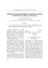
- 25728288, 2024, 1, Downloaded from https://onlinelibrary.wiley.com/doi/10.1002/vjch.202300059 by Readcube (Labtiva Inc.), Wiley Online Library on [01/05/2024]. See the Terms and Conditions (https://onlinelibrary.wiley.com/terms-and-conditions) on Wiley Online Library for rules of use; OA articles are governed by the applicable Creative Commons License LE ET AL. 39 FIGURE 1 The chemical structures compounds 1–5. (S)-(+)-Rhododenol (3): HRESIMS: m/z 189.0889 [M+Na]+ mined by measuring the absorbance at 405 nm. For quan- (C10 H14 O2 Na+ , 189.0891). [α]25 D +35.0 (CHCl3 , c 5.0×10⁻³ ). tification, one unit of α-glucosidase activity was defined as 1 H NMR (500 MHz, CDCl ): δ (J in Hz) 1.22 (3H, d, 6.5, H-4′), the amount of enzyme that liberated p-nitrophenol (1.0 μM) 3 H 1.72 (2H, m, H-2′), 2.62 (2H, m, H-1′), 3.83 (1H, dq, 12.5, 6.5, per minute. The IC50 value was calculated, representing H-3′), 6.75 (2H, d, 8.5, H-2, H-6), 7.04 (2H, d, 8.5, H-3, H-5). the concentration of the α-glucosidase inhibitor required 13 C NMR (125 MHz, CDCl ): δ 154.2 (C-1), 133.9 (C-4), 129.5 to inhibit 50% of enzymatic activity. As a positive control, 3 C (C-3, C-5), 115.4 (C-2, C-6), 67.8 (C-3′), 41.1 (C-1′), 31.3 (C-2′), acarbose, a known α-glucosidase inhibitor, was used in the 23.6 (C-4′). experiment. (+)-erythro-N-Benzoyl-3-phenylisoserine methyl ester (4): HRESIMS: m/z 322.1058 [M+Na]+ (C17 H17 NO4 Na+ , 322.1055). [α]25 D +40.0 (MeOH, c 5.15×10⁻³ ). 1 H NMR 3 RESULTS AND DISCUSSION (600 MHz, DMSO-d6 ): δH (J in Hz) 3.53 (3H, s, 1-OCH3 ), 4.49 (1H, dd, 9.6, 6.6, H-2), 5.42 (1H, dd, 10.8, 6.6, H-3), 5.81 Compound 1 was obtained as a yellow amorphous (1H, d, 9.6, OH), 7.25−7.84 (10H, m), 8.65 (1H, d, NH). 13 C powder and showed a sodiated molecular HRESIMS (High- NMR (150 MHz, DMSO-d6 ): δC 172.4 (C-1), 166.2 (C-1′), resolution Electrospray Ionisation Mass Spectrometry) 139.6−127.1, 73.7 (C-2), 56.0 (C-3), 51.6 (1-OCH3 ). ion at m/z 603.1269 [M+Na]+ (calcd. for C33 H24 O10 Na+ , (−)-α-Conidendrin (5): HRESIMS: m/z 379.1160 [M+Na]+ 603.1267). The 1 H NMR spectrum displayed signals for (C20 H20 O6 Na+ , 379.1158). [α]25 D −65.0 (CHCl3 , c 2×10⁻³ ). one 1,3,4,5-tetrasubstituted aromatic moiety [δH 6.62 (d, 1 H NMR (500 MHz, CDCl ): δ (J in Hz) 2.54 (1H, m, H-3a), J = 2.4 Hz, H-6), 6.74 (d, J = 2.4 Hz, H-8)], one ABX spin 3 H 2.61 (1H, dd, 13.5, 4.0, H-9a), 2.95 (1H, dd, 15.0, 10.5, H-9ax), system assignable to the 1,3,4-trisubstituted benzene rings 3.17 (1H, dd, 15.0, 4.0, H-9eq), 3.80 (3H, s, 3′-OCH3 ), 3.83 (1H, [δH 7.46 (d, J = 8.8 Hz, H-5′), 8.24 (dd, J = 8.8, 2.5 Hz, H-6′), d, 12.0, H-4), 3.86 (3H, s, 7-OCH3 ), 4.00 (1H, dd, 10.5, 8.5, H- 8.53 (d, J = 2.5 Hz, H-2′)] and one methine aromatic proton 3ax), 4.22 (1H, dd, 8.5, 6.5, H-3eq), 6.35 (1H, s, H-5), 6.58 (1H, [δH 7.03 (s, H-6″)]. Besides, two olefinic protons [δH 7.04 s, H-2′), 6.64 (1H, d, 8.0, H-6′), 6.67 (1H, s, H-8), 6.81 (1H, d, 8.0, (s, H-3), 7.17 (s, H-3″)] along with a pair of doublet signal H-5′). 13 C NMR (125 MHz, CDCl3 ): δC 178.4 (C-1), 148.0 (C-3′), [δH 7.06 (d, J = 8.5 Hz, H-3‴, H-5‴), 7.80 (d, J = 8.5 Hz, 146.5 (C-7), 144.8 (C-6), 145.4 (C-4), 134.3 (C-1′), 131.9 (C-8a), H-2‴, H-6‴)] of two hydrogens at ortho-positions of the 126.2 (C-4a), 121.6 (C-6′), 116.0 (C-5), 115.4 (C-5′), 112.1 (C- aromatic ring and three methoxy groups [δH 3.68 (s, 4‴- 8), 111.3 (C-2′), 72.5 (C-3), 56.1 (7-OCH3 ), 56.0 (3′-OCH3 ), 50.0 OCH3 ), 3.78 (s, 4′-OCH3 ), 3.88 (s, 7-OCH3 )] were observed (C-4), 47.8 (C-3a), 42.2 (C-9a), 29.3 (C-9). in the 1 H NMR spectrum. The 13 C NMR spectrum showed 33 carbons, including two carbonyl carbons (δC 183.0, 182.9), two olefinic carbons (δC 104.7, 104.1), and three 2.4 α-Glucosidase inhibitory assay methoxy groups (δC 56.0, 55.9, 55.4). Moreover, the signals of oxygenated carbons and aromatic carbons were found The α-glucosidase inhibitory activity was assessed follow- at δC 156.0–165.8 and 92.9–131.9 ppm, respectively. Based ing the modified method outlined by Kim et al.6 In this on the 1 H and 13 C NMR spectrum, compound 1 was char- assay, a mixture of 3 nM p-nitrophenyl-α-D-glucopyranoside acteristic of the biflavonoids with two flavone units. The (0.01 mL) and 20 U/mL α-glucosidase (0.01 mL) in 0.01 M HMBC (Heteronuclear Multiple Bond Correlation) correla- phosphate buffer (pH = 7) was combined with the sample tions between H-6, H-8, 7-OCH3 /C-7, H-5′, 4′-OCH3 /C-4′ solution (2.2 mL) to initiate the reaction. Each reaction was and H-2‴, H-3‴, H-5‴, H-6‴, 4‴- OCH3 /C-4‴ suggested the conducted at 37 ◦ C for 30 min and then halted by adding position of three methoxy groups at carbon C-7, C-4′, and 2 mL of 0.1 M Na2 CO3 . The enzymatic activity was deter- C-4‴, respectively. Additionally, three hydroxyl groups were
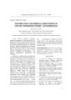
- 25728288, 2024, 1, Downloaded from https://onlinelibrary.wiley.com/doi/10.1002/vjch.202300059 by Readcube (Labtiva Inc.), Wiley Online Library on [01/05/2024]. See the Terms and Conditions (https://onlinelibrary.wiley.com/terms-and-conditions) on Wiley Online Library for rules of use; OA articles are governed by the applicable Creative Commons License 40 LE ET AL. FIGURE 2 Key HMBC correlations of compounds 1, 2, 5. determined at carbon C-5, C-5″, C-7″ based on the HMBC 3.83, dq, J = 12.5, 6.5 Hz) demonstrated the presence of 3- correlations between H-6/C-5, H-6″/C-5″ and H-6″/C-7″. hydroxybutanoyl moiety. The chemical structure of 3 was The C-3′-C-8″ linkage between two flavone units was determined as rhododenol because its NMR data were com- confirmed by observing the HMBC correlations between patible with those in the literature.9 The specific rotation H-2′/C-3′, H-2′/C-8″, and H-6″/C-8″. Thus, the structure of of 3 was [α]25 D +35.0 (CHCl3 , c 5.0 × 10⁻³ ); therefore, the 1 was assigned as sciadopitysin by comparing these data chemical structure of 3 was (S)-(+)-rhododenol.9 with the reported literature.7 Compound 4 was obtained as a white amorphous pow- Compound 2 was obtained as a yellow amorphous der and had the molecular formula C17 H17 NO4 through its powder. The molecular formula was determined to be HRESIMS ion at m/z 322.1058 [M+Na]+ (C17 H17 NO4 Na+ , C32 H22 O10 based on its HRESIMS ion at m/z 589.1115 322.1055). The 1 H NMR spectrum displayed the signals [M+Na]+ (calcd. for C32 H22 O10 Na+ , 589.1111). The 1 H and of one amide proton [δH 8.65 (d, J = 10.8 Hz, NH)], two 13 C NMR spectra were the same as those of 1, except for the methine protons [δH 4.49 (dd, J = 9.6, 6.6 Hz, H-2), 5.42 disappearance of one methoxy group. Therefore, the skele- (dd, J = 10.8, 6.6 Hz, H-3)], one hydroxy group [δH 5.81 (d, ton of 2 was confirmed as the biflavonoids with two flavone J = 9.6 Hz, OH)], one methoxy group [δH 3.53 (s, OCH3 )], units. The location of methoxy groups were proposed at car- and aromatic protons from δH 7.25 to 7.84 ppm. The 13 C bon C-7 and C-4′ due to the HMBC correlations between NMR spectrum showed the signals of two carbonyl car- H-6,H-8,7-OCH3 /C-7 and H-2′,H-5′,H-6′,4′-OCH3 /C-4′. Oth- bons (δC 172.4, 166.2), one oxygenated carbon (δC 73.7), erwise, the addition of HMBC correlations between H-2‴, one methine carbon (δC 56.0), one methoxy group (δC H-3‴, H-5‴, H-6‴/C-4‴ along with the available HMBC cor- 51.6), and other signals of two phenyl groups from 139.6 relations, which are similar to those of 1, four hydroxy to 127.1 ppm. Besides, the chemical shifts and splitting groups were suggested as C-5, C-5″, C-7″, and C-4‴. Two patterns of an amide group (δH 8.65, d, 10.8), methine flavone units were linked by the bridge between C-3′ and C- groups (δH 4.49, dd, 9.6, 6.6; 5.42, dd, 10.8, 6.6), a hydroxyl 8″ because of the observed HMBC correlations in Figure 2. group (δH 5.81, d, 9.6) and an aliphatic methoxy group (δH Based on all the aforementioned analyses, comparing the 3.53, s) proved the presence of a 3-phenylisoserine methyl NMR data of 2 with the reported literature data,8 compound ester fragment. Another phenyl group was attached directly 2 was identified as ginkgetin. to that fragment through the amide bridge. Therefore, Compound 3 was obtained as a white amorphous pow- the chemical structure of 4 was identified as N-benzoyl- der and indicated a sodiated molecular HRESIMS ion at 3-phenylisoserine methyl ester by comparing with the m/z 189.0889 [M+Na]+ (calcd. for C10 H14 O2 Na+ , 189.0891). literature.10 Moreover, the coupling constant determined The 1 H NMR spectrum of 3 exhibited the double signals the relative configuration of H-2 and H-3. Specifically, if of four protons at ortho-positions [δH 6.75 (d, J = 8.5 Hz, JH-2/H-3 = 3.5 Hz, both protons would be at the threo con- H-2, H-6), 7.04 (d, J = 8.5 Hz, H-3, H-5)], which proved the figuration and, inversely, at the erythro configuration.10 formation of AA′BB′ spin systems, two methylene protons Otherwise, JH-2/H-3 of 4 was 6.6 Hz; thus, the structure of 4 [δH 1.72 (m, H-2′), 2.62 (m, H-1′)], one methine proton [δH was erythro-N-benzoyl-3-phenylisoserine methyl ester. The 3.83 (dq, J = 12.5, 6.5 Hz, H-3′)] and one methyl group specific rotation of 4 was [α]25 D +40.0 (MeOH, c 5.15×10⁻³ ); [δH 1.22 (d, J = 6.5 Hz, H-4′)]. Besides, the 13 C NMR spec- therefore, the chemical structure of 4 was concluded as (+)- trum of 3 showed 10 carbons, including a methyl group erythro-N-benzoyl-3-phenylisoserine methyl ester.11 In the (δC 23.6), one oxygenated carbon (δC 67.8), two methylene experimental procedure, methanol was used as a common carbons (δC 41.1, 31.3), and the signals of aromatic carbon solvent. Therefore, although an acid catalyst has not been from 115.4 to 154.2 ppm. The splitting patterns of a methyl used during reflux and throughout the isolation process, group (δH 1.22, d, J = 6.5 Hz) and a methine proton (δH the possibility of 4 being an artifact cannot be ignored.
- 25728288, 2024, 1, Downloaded from https://onlinelibrary.wiley.com/doi/10.1002/vjch.202300059 by Readcube (Labtiva Inc.), Wiley Online Library on [01/05/2024]. See the Terms and Conditions (https://onlinelibrary.wiley.com/terms-and-conditions) on Wiley Online Library for rules of use; OA articles are governed by the applicable Creative Commons License LE ET AL. 41 TA B L E 1 α-Glucosidase inhibitory of isolated compounds. Inhibition (I%) Compound 250 µm 100 µm 50 µm 25 µm 10 µm IC50 (µm) 1 15.9 ± 1.6 3.69 ± 0.23 – – – >250 2 76.6 ± 2.1 50.3 ± 2.5 23.5 ± 1.3 5.2 ± 1.6 – 98.5 3 30.2 ± 1.6 12.0 ± 1.3 – – – >250 4 25.6 ± 1.2 7.3 ± 1.6 – – – >250 5 63.0 ± 1.8 37.9 ± 1.7 11.7 ± 1.6 – – 172.1 Acarbosea 64.5 ± 1.3 37.9 ± 1.2 15.5 ± 3.1 2.8 ± 1.7 – 168.0 –: Not shown inhibitory activity. a Positive control. However, methylation is a widely common functionaliza- The α-glucosidase inhibitory activity of the isolated tion method in the biosynthesis of natural products. There- compounds was evaluated and recorded in Table 1. The fore, this is more difficult to recognize a possible artifact.12 assay involved testing the compounds at various concen- Compound 5 was isolated as a white amorphous pow- trations, ranging from 10 to 250 μM. Among the tested der. Its molecular formula was identified as C20 H20 O6 based compounds, ginkgetin (2) and (−)-α-conidendrin (5) exhib- on its HRESIMS ion at m/z 379.1160 [M+Na]+ (calcd. for ited relatively weak α-glucosidase inhibitory activity in a C20 H20 O6 Na+ , 379.1158). The 1 H NMR spectrum exhibited concentration-dependent manner with the IC50 values of distinct signals corresponding to a 1,3,4-trisubstituted aro- 98.5 and 172.1 μM, respectively. matic moiety [δH 6.58 (s, H-2′), 6.64 (d, J = 8.0 Hz, H-6′), and 6.81 (d, J = 8.0 Hz, H-5′)], as well as a 1,2,4,5-tetrasubstituted aromatic moiety [δH 6.35 (s, H-5), and 6.67 (s, H-8)]. Addi- 4 CONCLUSION tionally, two methoxy groups were observed as singlets at δH 3.80 (s, 3′-OCH3 ), and 3.86 (s, 7-OCH3 ). Furthermore, the Five known compounds were isolated from the CHCl3 - spectrum displayed signals for an oxymethylene [δH 4.00 soluble fraction of the leaves of Taxus wallichiana (dd, J = 10.5, 8.5 Hz, H-3ax), 4.22 (dd, J = 8.5, 6.5 Hz, H- (Taxaceae), with the first report on separating compounds 3eq)], a bisbenzylic methine proton doublet [δH 3.83 (d, 1–4 from this plant. Among the isolated compounds, J = 12.0 Hz, H-4)], a benzylic methylene group [δH 2.95 (dd, ginkgetin (2) and (−)-α-conidendrin (5) showed weak J = 15.0, 10.5 Hz, H-9ax), 3.17 (dd, J = 15.0, 4.0 Hz, H-9eq)], α-glucosidase inhibitory activity. These results suggested and two methine protons [δH 2.54 (m, H-3a), and 2.61 (dd, that the use of Taxus wallichiana for treating diabetes in J = 13.5, 4.0 Hz, H-9a)]. The 13 C NMR spectrum displayed traditional medicines may be due to the α-glucosidase resonances corresponding to twelve aromatic carbons (δC inhibitory activity of phenolic compounds. 148.0-111.3), two aromatic methoxy carbons (δC 56.1, 56.0), ACKNOWLEDGMENTS an oxymethylene (δC 72.5), three methines (δC 42.2, 47.8, This research was funded by Vietnam National Univer- 50.0), and a methylene carbon (δC 29.3). Based on the sity, Ho Chi Minh City (VNU-HCM) under grant number observed HMBC correlations shown in Figure 2, the assign- 562-2022-18-04. ment of the 2,7′-cyclolignan structural feature was possible. Furthermore, the HMBC correlations between H-3/C-1, C- C O N F L I C T O F I N T E R E S T S TAT E M E N T 9a, C-3a, H-9a/C-1, C-3a, and H-3a/C-3, C-9a confirmed No potential conflict of interest was reported by the the presence of one γ-lactone unit attached to the 2,7′- authors. cyclolignan fragment via two methine carbons, C-9a and C-3a. The locations of two methoxy groups were suggested REFERENCES to be at C-7 and C-3′ from the HMBC correlation (Figure 2). 1. T. Mulliken, P. Crofton. Review of the Status, Harvest, Trade and Manage- Additionally, the coupling constant J confirmed the relative ment of Seven Asian CITES-Listed Medicinal and Aromatic Plant Species, configurations of H-4, H-3a, and H-9a. These protons would Bundesamt für Naturschutz, Bonn 2008. be at trans configuration if JH-9a/H-3a ≈ JH-4/H-3a ≥ 12.0 Hz, 2. H. Sharma, M. Garg. A review of traditional use, phytoconstituents and biological activities of Himalayan yew, Taxus wallichiana, J. Integr. and if JH-9a/H-3a ≈ JH-4/H-3a ≈ 9.0 Hz, they would be at Med. 2015, 13, 80. cis configuration.13 In compound 5, JH-9a/H-3a and JH-4/H-3a 3. J. L. McLaughlin, R. W. Miller, R. G. Powell, C. R. Smith Jr. were 13.5 and 12.0 Hz, respectively; these protons were 19-Hydroxybaccatin III, 10-deacetylcephalomannine and 10- concluded at trans-orientation. Finally, by comparing the deacetyltaxol: New antitumor taxanes from Taxus wallichiana, J. Nat. literature,14 the chemical structure of compound 5 was α- Prod. 1981, 44, 312. 4. P. H. Dang, H. X. Nguyen, T. T. T. Duong, T. K. T. Tran, P. T. Nguyen, T. K. T. conidendrin. The specific optical rotation of 5 was [α]25 D Vu, H. C. Vuong, N. H. T. Phan, M. T. T. Nguyen, N. T. Nguyen, S. Awale. −65.0 (CHCl3 , c 2 × 10⁻³ ), therefore, the chemical structure α-Glucosidase inhibitory and cytotoxic taxane diterpenoids from the of 5 was concluded as (−)-α-conidendrin.14 stem bark of Taxus wallichiana, J. Nat. Prod. 2017, 80, 1087.
- 25728288, 2024, 1, Downloaded from https://onlinelibrary.wiley.com/doi/10.1002/vjch.202300059 by Readcube (Labtiva Inc.), Wiley Online Library on [01/05/2024]. See the Terms and Conditions (https://onlinelibrary.wiley.com/terms-and-conditions) on Wiley Online Library for rules of use; OA articles are governed by the applicable Creative Commons License 42 LE ET AL. 5. P. H. Dang, H. X. Nguyen, H. H. T. Nguyen, T. D. Vo, T. H. Le, T. H. N. 12. A. Venditti. What is and what should never be: Artifacts, improbable Phan, M. T. T. Nguyen, N. T. Nguyen. Lignans from the roots of Taxus phytochemicals, contaminants and natural products, Nat. Prod. Res. wallichiana and their α-glucosidase inhibitory activities, J. Nat. Prod. 2020, 34, 1014. 2017, 80, 1876. 13. R. Dhal, Y. Nabi, E. Brown. Etudes de lignanes 7-syntheses totales 6. K. Y. Kim, K. A. Nam, H. Kurihara, S. M. Kim. Potent α-glucosidase des (±)-α-et-β-conidendrines et des methyl (±)-α-et-β-conidendrals, inhibitors purified from the red alga Grateloupia elliptica, Phytochem- Tetrahedron 1986, 42, 2005. istry 2018, 69, 2820. 14. J. Fischer, A. J. Reynolds, L. A. Sharp, M. S. Sherburn. Radical car- 7. S. H. Li, H. J. Zhang, X. M. Niu, P. Yao, H. D. Sun, H. H. S. Fong. boxyarylation approach to lignans. Total synthesis of (−)-arctigenin, Chemical constituents from Amentotaxus yunnanensis and Torreya (−)-matairesinol, and related natural products, Org. Lett. 2004, 6, yunnanensis, J. Nat. Prod. 2003, 66, 1002. 1345. 8. Y. Konda, T. Sasaki, H. Kagawa, H. Takayanagi, Y. Harigaya, X. L. Sun, X. Li, M. Onda. Conformational analysis of C3′-C8 connected biflavones, J. Heterocycl. Chem. 1995, 32, 1531. 9. Y. Yuasa, S. Shibuya, Y. Yuasa. Resolution of racemic rhododendrol by lipase-catalyzed enantioselective acetylation, Synth. Commun. 2003, How to cite this article: T. D. Le, D. V. T. Le, T. H. 33, 1469. Tran, P. H. Dang, T. H. Le, M. T. T. Nguyen, N. T. 10. Z. Zhou, X. Mei. A practical and stereoselective synthesis of taxol side Nguyen. Chemical constituents of the leaves of chain, Synth. Commun 2003, 33, 723. 11. S. Jost, Y. Gimbert, A. E. Greene, F. Fotiadu. Totally stereocontrolled Taxus wallichiana (Taxaceae), Vietnam J. Chem. 2024, nitrone-ketene acetal based synthesis of (2S,3S)-N-benzoyl- and 62, 37. https://doi.org/10.1002/vjch.202300059 N-boc-phenylisoserine, J. Org. Chem. 1997, 62, 6672.
CÓ THỂ BẠN MUỐN DOWNLOAD
-

Two long chain alkyl alcohols from the leaves of acroton tonkinensis gagnep, euphorbiaceae
 1 p |
1 p |  35
|
35
|  5
5
-
A new diterpene glycoside from the stem bark of acanthopanax trifoliatus
4 p | 53
| 3
-

Tovopyrifolin C and Amentoflavone from the leaves of the vietnamese plant Calophyllum inophyllum L.
2 p | 33
| 3
-
Apigenin 7-O-B-glucoside from the leaves of Acanthus integrifolius T. Anders., Acanthaceae
3 p | 65
| 3
-
Further Study on Chemical Constituents of Croton tonkinensis Gagnep., Euphorbiaceae
2 p | 56
| 3
-
Chemical constituents of the Moringa oleifera LAM. leaves collected in Bac Ninh province
5 p | 27
| 3
-
Chemical constituents of ethyl acetate extract from Taxus wallichiana ZUCC. (taxaceae) leaves collected in Ha Giang province
6 p | 25
| 2


Chịu trách nhiệm nội dung:
Nguyễn Công Hà - Giám đốc Công ty TNHH TÀI LIỆU TRỰC TUYẾN VI NA
LIÊN HỆ
Địa chỉ: P402, 54A Nơ Trang Long, Phường 14, Q.Bình Thạnh, TP.HCM
Hotline: 093 303 0098
Email: support@tailieu.vn
Giấy phép Mạng Xã Hội số: 670/GP-BTTTT cấp ngày 30/11/2015 Copyright © 2022-2032 TaiLieu.VN. All rights reserved.

