Giant duodenal lipoma
-
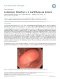
A 55-year-old woman presented with a 3-year history of gradually progressive postprandial epigastric fullness. Esophagogastroduodenoscopy revealed a large, broad-based mass in the medial wall of the duodenal bulb (Figure 1). Endoscopic ultrasound showed a large, hyperechoic lesion originating from the submucosal layer of the duodenal wall consistent with a lipoma, and computed tomography of the abdomen with contrast suggested the presence of a homogeneous mass with uniform fat density in the duodenal bulb (Figure 2).
 3p
3p  covid19
 11-05-2020
 19
19
 1
1
 Download
CHỦ ĐỀ BẠN MUỐN TÌM


