


Figure Drawing - Individual Muscles - Rear Limb
lượt xem 12
download
 Download
Vui lòng tải xuống để xem tài liệu đầy đủ
Download
Vui lòng tải xuống để xem tài liệu đầy đủ
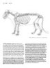
Gluteus superficialis HORSE • Origin: Point of the hip (coxal tuberosity) and an adjacent area on the outer edge of the ilium of the pelvis; from fascia covering the gluteus medius (in part ultimately originating from the ligament connecting the sacrum to the ilium). • Insertion: Third trochanter of the femur, one third of the way down the outside of the bone. • Action: Flexes the hip joint; pulls the limb away from the body. • Structure: The gluteus superficialis is a thin, V-shaped muscle that converges on the femur. The front portion is partly covered by, and firmly...
Bình luận(0) Đăng nhập để gửi bình luận!
Nội dung Text: Figure Drawing - Individual Muscles - Rear Limb
- 88 INDIVIDUAL MUSCLES » REAR LIMB HORSE DOG Gluteus superficialis DOG AND FELINE HORSE • Origin: The sacrum, the first tail vertebra, the front half of the ligament • Origin: Point of the hip (coxal tuberosity) and an adjacent area on the connecting the sacrum to the ischiatic tuberosity (sacrotuberal ligament), outer edge of the ilium of the pelvis; from fascia covering the gluteus and the fascia covering the gluteus medius. medius (in part ultimately originating from the ligament connecting the • Insertion: Outer surface of the femur, about one-eighth of the way sacrum to the ilium). down the bone. • Insertion: Third trochanter of the femur, one third of the way down the • Structure: The gluteus superficialis is a small, flat muscle appearing outside of the bone. somewhat rectangular on the surface. It is smaller than the gluteus • Action: Flexes the hip joint; pulls the limb away from the body. medius. • Structure: The gluteus superficialis is a thin, V-shaped muscle that con- verges on the femur. The front portion is partly covered by, and firmly The gluteus superficialis is not present in the ox. The upper front portion attached to, the tensor fasciae latae muscle. The rear portion sits on top of the gluteobiceps muscle of the ox is believed to be the rear portion of of the gluteus medius; its rear edge is covered by the biceps femoris. the gluteus superficialis, and the rear portion of the tensor fasciae latae may be the front portion of the gluteus medius.
- INDIVIDUAL MUSCLES + REAR LIMB 89 LION Caudofemoralis (Gluteofemoralis) elongated triangular muscle located behind the gluteus superficialis. FELINE Approximately one third of the way down the thigh, it disappears under • Origin: Side projections of the first, second, and third tail vertebrae. the biceps femoris. About two thirds of the way down, it develops a long, • Insertion: The fascia of the leg in front of the biceps femoris muscle, very thin tendon. The visible superficial portion of the muscle belly is and the middle of the outside edge of the patella. approximately the same size as the gluteus superficialis. • Action: Extends the hip joint; pulls the limb away from the body. • Structure: The caudofemoralis muscle, exclusive to the felines, is an
- 90 INDIVIDUAL MUSCLES > REAR LIMB HORSE DOG Tensor fasciae latae the underlying vastus lateralis muscle. This can create a narrow form, HORSE directed from the point of the hip to the patella. • Origin: Point of the hip (outer front corner of the ilium of the pelvis). OX • Insertion: Into the fascia of the leg that surrounds the vastus lateralis • Structure: The lower end of the belly ends in a wide inverted "V." and the rectus femoris, therefore indirectly into the patella, the outer The front edge of the belly ends a short distance above the patella. patellar ligament, and the front edge of the tibia. DOG AND FELINE • Action: Flexes the hip joint and, by its ultimate attachment to the patel- • Origin: Lower edge of the front end of the pelvis; the surface of the la and the tibia, extends the knee joint. gluteus medius. • Structure: The tensor fasciae latae is a triangular muscle that forms the • Insertion: Into the fascia covering the thigh muscles. front edge of the upper end of the thigh. Its belly begins on the point of • Structure: The triangular muscle separates into two forms on the the hip and ends midway between the point of the hip and the patella. surface. The muscle belly ends high on the thigh; its lower edge Its rear edge tightly adheres to the gluteus superficialis. The muscle is directed downward and forward from the upper end of the femur. belly may separate into two forms upon contraction. The sartorius, not the tensor fasciae latae, is the leading muscle A thickened, narrow band of fascia coming off the belly passes on the front of the thigh. over the thigh muscles and attaches to the patellar ligament. When the tensor fasciae latae is tensed, the fascial band tightens and compresses
- INDIVIDUAL MUSCLES + REAR LIMB 91 DOG Sartorius • Action: Flexes the hip joint; pulls the limb toward the centerline of HORSE AND OX the body. The sartorius is a minor muscle and is rarely visible on the surface. It is • Structure: The sartorius begins on the front end of pelvis and ends on a small, narrow muscle that lies on the inside of the thigh, just in front of the inside of the knee. It passes down the front and inside of the thigh, the gracilis. Originating deep on the fascia and tendon in the region veering to the inside of the knee and becoming a wide tendon before where the upper inner end of the thigh meets the rear of the abdomen, inserting. In the side view of the body, the front edge of the sartorius can it becomes tendinous above the knee. Its tendon ultimately inserts into be seen passing down most of the front of the thigh and disappearing as the medial patellar ligament and the tibia. it shifts to the inside. The lower half of the muscle passes over the lower end of the rectus femoris and the vastus medialis, adding muscular DOG AND FELINE thickness on the lower end of the inside of the thigh. The muscle can • Origin: Front portion: Line on the front edge of the pelvis. Rear portion: also be seen in the front and inside views of the leg. Line on the lower edge of the front end of the pelvis. The sartorius consists of two elongated parallel muscular bands— • Insertion: Dog: Front portion: With the vastus medialis and the rectus a front and a rear portion—in the dog, but a single, wider muscle in femoris into the patella and the fascia of the knee. Rear portion: Front the feline. edge of the tibia. Feline: Continuous insertion from the patella to the upper end of the tibia.
- 92 INDIVIDUAL MUSCLES » REAR LIMB HORSE DOG Quadriceps femoris: Vastus lateralis, medialis, groove running down the front of the mass of the three vastus muscles and intermedius, Rectus femoris in which the rectus femoris sits. The rectus femoris is an elongated HORSE muscle, tapered at both ends. • Origin: Vastus lateralis: Outer surface of the femur, from a level just The patellar ligaments, although termed "ligaments" because below the hip socket to two thirds of the way down the bone. Vastus they connect bone to bone (the patella to the tibia), are actually a medialis: Inner surface of the femur, from a level just below the hip sock- continuation of the quadriceps muscle and are its tendons of insertion. et to two thirds of the way down the bone. Rectus femoris: Two small In the horse and the ox, three patellar ligaments—inner, middle, and adjacent areas on the body of the pelvis just in front of the hip socket. outer—converge on the tibial tuberosity. • Insertion: All parts: The entire front surface and upper edge of the OX patella, and because of the attachment of the three patellar ligaments to • Insertion: Vastus lateralis: Also into the lateral patellar ligament. the tibia, ultimately into the front of the upper end of the tibia (the tibial DOG AND FELINE tuberosity). The vastus lateralis and medialis also insert into the sides • Origin: Small areas on the inside (vastus medialis) and outside (vastus of the rectus femoris, attaching to the fascia covering its surface. In lateralis) of the femur near its upper end. The rectus femoris originates addition, the vastus medialis inserts into the upper half of the medial from a single area on the pelvis. patellar ligament. • Insertion: All parts into the patella, therefore ultimately into the tibia. • Action: All parts extend the knee joint; the rectus femoris also flexes The vastus muscles insert into their respective sides of the rectus the hip joint. femoris. • Structure: The quadriceps muscle consists of the vastus lateralis, the • Structure: The quadriceps femoris in the dog and feline does not vastus medialis, the deep vastus intermedius, and the rectus femoris. bulge forward as much as in the horse, but the vastus lateralis bulges The three vastus muscles all begin on the femur, and the rectus femoris out to the side quite a bit, especially at its upper end. Although it is originates on the pelvis. These four components form a large, wide partially covered by the biceps femoris, the tensor fasciae latae, and the (front-to-back) but flattened (side-to-side) muscle mass. It embraces the sartorius, the quadriceps femoris produces the bulk of the form on the inside, outside, and front of the femur, but it lies for the most part in front of the thigh. front of it. The vastus lateralis lies on the outside of the thigh and is There is a single patellar ligament between the patella and somewhat oval in outline. Its rear edge is straighter than the front edge, the tibia. and its rear portion is covered by the biceps femoris. The vastus medialis is similar in shape, and lies on the inside of the thigh. There is a wide
- INDIVIDUAL MUSCLES > REAR LIMB 93 OX Gluteobiceps front portion passing from the hip region to the knee, and a long trian- OX gular rear portion beginning at the rear end of the pelvis. The • Origin: Spines of the sacrum, the sacrotuberal ligament, the ischiatic division between these two portions is more visible near the pelvis, tuberosity at the rear end of the pelvis, and the fascia covering the and it diminishes lower down, where the muscle fibers attach to the leg gluteus medius and the tail. fascia. A long, narrow, tendinous band passes from the lower end of • Insertion: Into the fascia of the leg, ultimately into the patella, the the muscle to the heel. lateral patellar ligament, the front edge of the tibia, and the heel bone. The gluteobiceps consists of the biceps femoris fused to the rear • Action: Extends the hip joint; extends the ankle joint; pulls the limb portion of the gluteus superficialis. It is not present in the horse, dog, away from the body. With different portions, it both flexes and extends or feline, where there are separate gluteus superficialis and biceps the knee joint. femoris muscles. • Structure: The gluteobiceps is a very large, roughly rectangular mus- cle, wide above and below, and narrower in the middle. Its front edge is thin, and its rear edge is thick. It is divisible into two portions—a large
- 94 INDIVIDUAL MUSCLES + REAR LIMB Biceps femoris • Action: The entire muscle extends the hip joint. The upper front fibers HORSE extend the knee joint, and the lower rear fibers extend the ankle joint • Origin: Long head: From the ligament connecting the sacrum to the and may also flex the knee joint. ilium of the pelvis, in the vicinity of the third and fourth sacral spines, • Structure: Large, wide muscle that begins narrow at the pelvis and and from the surface of the gluteal muscles and the tail. Short head: then fans out to cover the rear portion of the outside of the thigh. Lower edge of the rear end of the pelvis. Because its muscle fibers insert into a wide sheet of fascia that ulti- • Insertion: All portions fuse deeply and then first insert into the back of mately inserts further on, contracting muscle fibers pull on the fascia the femur, about one third of the way down the bone. The lower ends of and may create raised ridges in line with the direction of those muscle the muscle develop into a wide aponeurotic (tendinous) sheet that fuses fibers. This pulling can distort the volumes of the underlying muscles. into the fascia of the leg at and below the knee. The two heads ultimate- The line where the muscle fibers attach to the fascia can occasionally ly insert as follows: Long head—into the patella and lateral patellar liga- be seen on the surface, which can also confuse the volumes of the ment. Short head/front portion—into the lateral patellar ligament and muscles of the leg. the front edge of the tibia. Short head/rear portion—into the leg fascia, Along the rear edge of the muscle, new fibers begin deeply, and the end of the heel bone (calcaneus). wrap around to the outside, and then pass downward and forward on • Action: Entire muscle extends the hip joint (forward propulsion, kick- the surface. This structure may divide the overall muscle into several ing, rearing), and pulls the limb away from the body. The long head subtle forms. extends the knee joint, the short head/front portion flexes the knee A long, narrow tendon develops on the deep surface of the biceps joint, and the short head/rear portion flexes the knee joint and extends femoris, emerges at its lower end, and descends along the surface of the the ankle joint (extends the foot). gastrocnemius muscle. It eventually fuses with the Achilles tendon, • Structure: The biceps femoris is a massive muscle that consists of a which attaches to the heel bone (calcaneus). long head (long vastus) and a short head that separates into two por- FELINE tions, producing a total of three forms. The long head is crescent- • Origin: Only from the ischial tuberosity, not from the sacrotuberal liga- shaped—widest at its center (where it covers the greater trochanter of ment. the femur) and tapered at its ends. It begins at the top of the sacrum and • Structure: Because of its small point of origin, the biceps femoris ends at the level of the bottom of the patella. Its front edge is thin, assumes a more fan-shaped belly, and its fiber arrangement is simpler, whereas its rear edge is thicker and more clearly defined on the surface. than it is in the dog. It is divisible into a large front portion and a smaller The short head is a triangular volume that separates into two forms. Its rear portion. front portion flattens and lies on the outside of the knee. The rear por- tion, which is thicker and rounder, ends distinctly on the surface of the The ox has no biceps femoris, but rather a gluteobiceps, which is an outer side of the gastrocnemius muscle. A long, narrow tendon passes extensive, complex, single-bellied muscle consisting of the combined from the lower end of the biceps femoris muscle to the heel. biceps femoris and gluteus superficialis. The semitendinosus muscle covers the rear edge of the upper half of the long head and the upper end of the short head. A strong vertical groove on the back of the thigh separates the biceps femoris from the semitendinosus. DOG • Origin: Superficial head: Outer corner of the ischiatic tuberosity at the rear end of the pelvis, and the rear third of the sacrotuberal ligament. Deep head: Bottom of the outer corner of ischiatic tuberosity, deep to the origin of the superficial portion. • Insertion: Into the fascia of the leg, ultimately into the patella, the patellar ligament, the front edge of the upper end of the tibia, and the end of the heel bone (calcaneus).
- INDIVIDUAL MUSCLES » REAR LIMB 95 HORSE DOG
- 96 INDIVIDUAL MUSCLES > REAR LIMB Semitendinosus DOG AND FELINE HORSE • Origin: Outer corner of the rear end of the pelvis. • Origin: First and second tail vertebrae, fascia of the tail, and the lower • Insertion: Front edge of the tibia, about one fourth of the way down the edge of the rear end of the pelvis. tibia, and the heel bone (calcaneus). • Insertion: Fascia of the inside of the leg, ultimately into the front edge • Structure: This is an elongated muscle that passes between the rear of the tibia and the heel bone (calcaneus). end of the pelvis and the upper end of the inside of the knee region. Its • Action: Extends the hip joint and the ankle joint; flexes the knee joint; lower end, flattened from side-to-side, sends off a wide tendon to the rotates the leg inward. front edge of the tibia and a long, narrow one to the heel. The muscle • Structure: The semitendinosus is a long muscle that begins on the top belly lies on the middle of the back of the thigh and forms a very small of the base of the tail, passes down the back of the thigh, and ends on part of the middle portion of the rear profile of the thigh. The semi- the inside of the upper end of the tibia. It forms the entire rear profile of membranosus forms the upper profile, and the biceps femoris creates the thigh in the horse. The muscle belly is triangular in cross section. the lower profile. The semitendinosus begins thin and narrow at the tail, and gets thicker The semitendinosus descends in contact with the biceps femoris as it descends. The lower end becomes thin again and flattened side-to- located to its outside. When they reach the back of the knee, the biceps side, then terminates in a wide tendon that fuses with the fascia of the femoris veers toward the outside (and descends lower), whereas the inside of the leg. A separate long, narrow, tendinous band passes from semitendinosus shifts inward. This leaves a triangular depression on the the lower end of the belly to the heel bone. The upper part (between the back of the knee. tail and the rear end of the pelvis) is unique to the horse. A deep head originates from the rear end of the pelvis, but it soon fuses to the main body of the muscle. OX • Origin: Only from the lower edge of the rear end of the pelvis. • Structure: The fleshy muscle is elongated and slightly tapered at both ends. Its lower end is flattened side-to-side. The semitendinosus forms the lower edge of the rear profile of the thigh; the semimembranosus projects past it to form the upper edge.
- INDIVIDUAL MUSCLES > REAR LIMB 97 HORSE OX DOG
- 98 INDIVIDUAL MUSCLES » REAR LIMB HORSE DOG Semimembranosus • Insertion: Inner surface of the lower end of the femur, and the inner HORSE surface of the uppermost end of the tibia. • Origin: Rear free edge of the sacrotuberal ligament (passing from the • Structure: The semimembranosus in the ox more closely resembles this second tail vertebra to the top of the rear end of the pelvis) and the muscle of the dog than the horse. The lower end splits and sends a lower edge of the rear end of the pelvis. major portion to the femur and a smaller portion to the tibia. This sepa- • Insertion: Inner surface of the lower end of the femur. ration and insertion is deep and not visible on the surface. • Action: Extends the hip joint; pulls the limb toward the centerline of The upper end of the belly slightly projects past the semitendi- the body. nosus to form the upper portion of the rear profile of the thigh. • Structure: The semimembranosus is a large, thick muscle located on DOG AND FELINE the rear portion of the inside of the thigh, where most of it comes to • Origin: A line on the lower edge of the rear end of the pelvis. the surface. It runs alongside the semitendinosus and is partly covered • Insertion: A vertical line on the inner back corner of the lower end of by the gracilis, mostly at its lower end. The muscle begins at the base the femur, ending a short distance from the bottom of the bone, and a of the tail and ends on the inside of the knee. Pointed on its upper small area on the inner side of the uppermost end of the tibia. end, the descending muscle belly is joined by a deep head that origi- • Structure: This is a thick fleshy muscle consisting of two heads. One nates from the rear end of the pelvis. The upper end of the muscle, head inserts into the femur, while the other crosses the knee joint and between the tail and the pelvis, is unique to the horse. The semimem- inserts into the tibia. The belly comes to the surface on the upper, inner branosus is not seen in the side view of the thigh (it does not form rear corner of the thigh, between the semitendinosus and the gracilis. part of the rear profile). The upper portion of the muscle projects past the semitendinosus to OX form the upper portion of the rear profile of the thigh. • Origin: From the lower edge of the rear end of the pelvis only, but from a more extensive area than in the horse.
- INDIVIDUAL MUSCLES > REAR LIMB 99 HORSE DOG Gracilis OX HORSE • Insertion: Also to the heel bone. • Origin: Front two thirds of the line effusion of the two halves of the • Action: Also extends the ankle joint. pelvis, on the mid line on the bottom of the pelvis (variously from bone, • Structure: The lower end of the muscle belly also sends a tendinous ligament, and tendon). band to the heel bone. • Insertion: Medial patellar ligament, inner surface of the tibia, and the DOG AND FELINE fascia of the leg. • Origin: Line on the midline on the bottom of the pelvis that veers • Action: Primarily pulls the limb toward the centerline of the body; outward as the rear projections of the pelvis diverge. extends the hip joint. • Insertion: Ultimately into the front edge of the tibia and into the • Structure: The gracilis is a wide, thin, somewhat rectangular muscle heel bone. lying on the rear portion of the inner side of the thigh. This flat muscle • Action: Also flexes the knee joint and extends the ankle joint. thins toward its rear edge. It shares its origin with the same muscle of • Structure: The muscle thickens toward its rear edge, opposite to that the other leg; the upper ends of the two muscles are in contact with of the horse. The muscle belly is also narrower than in the horse. In each other when the animals is in the standing position. The muscle addition to the tendon to the front of the tibia, the lower end of the belly belly ends below, developing a wide tendon that fuses with the fascia of sends a tendinous band back to the heel bone, along with the semi- the leg. tendinosus.
- 1OO INDIVIDUAL MUSCLES * REAR LIMB HORSE DOG Tibialis cranialis (Tibialis anterior in humans) on the lower row of tarsal bones and the upper end of the large HORSE metatarsal. It pierces the tendon of the peroneus tertius, as in the horse. • Origin: Upper end of the outer side of the tibia, including the concavity DOG AND FELINE and adjacent bony prominences, and the fascia of the leg. • Origin: Vertical line on outer side of tibia, just to the outside of the • Insertion: Front of the upper end of the large metatarsal bone, and the front edge, continuing up into the concavity at the top of the outside of inside back corner of the ankle (into the rear bone on the lower tarsal row). the bone. Feline: Also into the upper end of the fibula. • Action: Flexes the ankle joint. • Insertion: Dog: Inner edge of the foot, into the lower tarsal bone and • Structure: The tibialis cranialis is a flattened muscle lying on the front the adjacent upper end of the inner metatarsal. Feline: Into the of the tibia. Its flat face is directed forward and outward; the extensor metatarsal only. digitorum longus and the peroneus tertius lie on this surface. The mus- • Action: Flexes the ankle joint; rotates the foot slightly outward. cle begins fleshy above and becomes tendinous below. In front of the • Structure: This elongated muscle is wider at its upper end. In the dog, ankle, its tendon emerges through the perforated tendon of the overly- it develops a flat tendon two thirds of the way down the bone. In the ing peroneus tertius, and then splits, sending one branch straight down feline, the lower end of the muscle belly becomes tendinous closer to and the other around to the inside of the ankle. the ankle than in the dog, covering more of the belly of the extensor Only the inner edge of the muscle belly is visible on the surface digitorum longus. The tendon descends and then veers inward, cross- between the tibia and the extensor digitorum longus. The inner branch ing the front of the foot, and then ends on the inner edge of the foot. of the tendon can be seen when the ankle is flexed. The slightly flattened belly sits on the outer front edge of the lower leg OX and wraps (side-to-side) around the underlying muscles. The tibialis • Structure: The tibialis cranialis is a thinner and more concealed muscle cranialis is not covered by the extensor digitorum longus and the than in the horse. Only the upper end of the belly, between the tibia and peroneus tertius, as it is in the horse and the ox. the peroneus tertius, and the tendon, is superficial. The tendon remains single and veers to the inside of the ankle to attach into adjacent areas
- INDIVIDUAL MUSCLES » REAR LIMB 101 HORSE OX Peroneus tertius (Fibularis tertius) The peroneus tertius has virtually no effect on the surface, but is HORSE presented here because its lower end is superficial, and for comparison • Origin: From a depression on the outer surface of the very bottom of with the same muscle of the ox. the femur. OX • Insertion: Upper front end of the large metatarsal bone and the front of • Insertion: Inner front corner of the upper end of the large (fused) the tarsal bone above it; the outside of the ankle, into the base of the metatarsal; inner rear corner of the ankle, into several adjacent bones, heel bone and the adjacent tarsal bone just below it. including the upper end of the large metatarsal bone. • Action: Rigid cable that forces the ankle joint to flex when the knee • Action: Flexes the ankle joint. joint is flexed. • Structure: The muscle has a fleshy, slightly flattened belly, rather than • Structure: The peroneus tertius is a strong tendon, devoid of any mus- just a tendon, as in the horse. It lies superficially on the front of the cle fibers, which begins on the femur and ends on the ankle. lower leg. Passing from the knee to the ankle, it begins and ends with Sandwiched between the bellies of the tibialis cranialis and the extensor tendons; its muscle belly is pointed at both ends. At the level of the digitorum longus on the front of the lower leg, it emerges from between lower end of the tibia, the tendon is perforated, allowing the tendon of them approximately two thirds of the way down the leg. At the level of the underlying tibialis cranialis to emerge, as in the horse. Below the the bottom of the tibia, the peroneus tertius is perforated, and the ten- perforation, it sends a tendinous branch to the inside of the ankle (oppo- don of the underlying tibialis cranialis emerges through this hole. The site that of the horse). The peroneus tertius covers most of the extensor peroneus tertius then continues straight down to the metatarsal bone. digitorum longus and is closely adherent to it. Below the level of the perforation, it sends off a small tendinous branch that passes around to the outside of the ankle, splits, and then attaches The peroneus tertius is not present in the dog or the feline; the extensor to the outside of the ankle. digitorum lateralis has in the past been called the peroneus tertius.
- 102 INDIVIDUAL MUSCLES > REAR LIMB OX HORSE DOG Extensor digitorum longus (Extensor pedis) peroneus tertius. The muscle, overall, is pointed at both ends and con- HORSE sists of two bellies, each of which develops a long tendon at the level of • Origin: From a depression on the outer surface of the very bottom of the ankle joint. The inner tendon inserts into the inner toe. The outer the femur, in common with the tendon of the peroneus tertius. tendon splits at its lower end and inserts into both toes. • Insertion: Upper edge of the front surface of all three toe bones, prima- DOG AND FELINE rily into the last. • Insertion: Last toe bone of all four digits. • Action: Extends all three toe joints; flexes the ankle joint. • Structure: The upper end of the belly of the extensor digitorum longus • Structure: The extensor digitorum longus is a long muscle with a very is covered by the tibialis cranialis and the peroneus longus. The extensor long tendon that passes from the femur to the last toe. The slightly digitorum longus then emerges from between them, one third of the way flattened muscle belly, tapered at both ends, lies on the outer front cor- down the tibia. It becomes tendinous before reaching the ankle joint, ner of the lower leg. It becomes tendinous above the level of the ankle and then travels alongside the tendon of the tibialis cranialis for a short joint. The long tendon lies on the front of the leg bones and disappears distance. The tendon of the tibialis cranialis soon moves away, veering to under the hoof. Halfway down the metatarsal bone, the tendon is the inside of the ankle. The tendon of the extensor digitorum longus joined by the tendon of the extensor digitorum lateralis. Lower down, then separates into four tendons, one for each toe. on the upper toe bone, both edges of the tendon receive branches from In the feline, the belly of the tibialis cranialis is wider and the suspensory ligament. descends lower than in the dog, thereby covering more of the extensor OX digitorum longus, which closely adheres to it. This leaves the outer part • Insertion: Upper edge of the front surface of the last toe bone of both of the lower portion of the extensor digitorum longus visible on the sur- toes, and the upper edge of the middle toe bone of the inner toe. face. The muscle fibers of the belly continue down to the level of the • Structure: The muscle is much thinner than in the horse, and also more ankle joint before it becomes tendinous. concealed, as its inner portion is covered by, and closely adheres to, the
- INDIVIDUAL MUSCLES » REAR LIMB 103 HORSE OX DOG Extensor digitorum lateralis • Insertion: Upper front end of the middle toe bone of the outer toe. HORSE (Peroneus) • Action: Extends the upper two toe joints of the outer toe. • Origin: Outer surface of the upper end of the tibia, outer surface of the • Structure: Similar to that in the horse, but the tendon continues inde- fibula, wide ligament between the tibia and the fibula, and the ligament pendently all the way down to the toe. on the outside of the knee joint between the femur and the fibula. DOG AND FELINE • Insertion: Into the tendon of the extensor digitorum longus, one third • Origin: Dog: Front surface of the upper third of the shaft of the fibula. of the way down the metatarsal, therefore ultimately into the three Feline: Outer surface of the upper half of the shaft of the fibula. toe bones. • Insertion: Into the tendon of the extensor digitorum longus to the out- • Action: Assists the extensor digitorum longus in extending the ermost toe, therefore ultimately into the last toe bone. toe bones. • Action: Extends the toe bones of the outermost toe; pulls the outer- • Structure: The extensor digitorum lateralis has an elongated, flattened most toe away from the foot. belly located on the outside of the lower leg. The belly begins at the • Structure: The extensor digitorum lateralis is a small muscle lying deep level of the knee joint and becomes tendinous at the lower end of the in the lower leg. In the dog, only its tendon comes to the surface on the tibia. The tendon passes through a shallow groove on the outside of the lower half of the outer side of the lower leg, where it lies between the lower end of the tibia, passes over the spool of the adjacent tarsal bone, tendons of the peroneus longus in front and the peroneus brevis behind. and then angles forward. It veers inwardly in its descent and then In the feline, some of the lower part of the muscular belly can be seen merges with the tendon of the extensor digitorum longus. on the surface. The tendon hooks behind the lower end of the fibula, OX along with, but in front of, the tendon of the peroneus brevis. It passes • Origin: Outer surfaces of the upper end of the tibia and the vestigial under the tendon of the peroneus longus, and then descends along the head of the fibula, and the ligament on the outside of the knee between outer edge of the front surface of the foot. It merges into the tendon of the femur and the tibia. the extensor digitorum longus on top of the upper toe bone.
- 104 INDIVIDUAL MUSCLES > REAR LIMB OX DOG Peroneus longus (Fibularis longus) • Action: Rotates the foot, directing the rear surface outward. OX • Structure: The peroneus longus consists of a short belly lying on the • Origin: Outer surface of the upper end of the tibia, and the ligament upper half of the outside of the lower leg and a long tendon that reaches connecting the femur to the tibia. the foot. The upper end of the belly, along with the tibialis cranialis • Insertion: Upper end of the inner rear corner of the large metatarsal which it touches, covers the upper portion of the extensor digitorum bone and the adjacent tarsal bone above it. longus. Its long tendon passes over the outside of the fibula, in its own • Action: Flexes the ankle joint; rotates the foot inwardly. groove, angles forward slightly, descends to the level of the upper end of • Structure: The muscle belly forms a narrow, elongated, inverted trian- the metatarsals, and then sharply curves around to the back of the foot gle that lies on the upper half of the outside of the lower leg. The belly to insert in a horizontal line across the tops of the metatarsals. begins wide at its origin, and it then tapers before becoming tendinous In the feline, the peroneus longus becomes tendinous two thirds halfway down the tibia. The tendon passes over the outside of the ankle of the way down the fibula. and then curves around the back of the foot to insert on its inner rear corner. In the horse, the muscle formerly called the peroneus is not a true per- oneus muscle, but is correctly called the extensor digitorum lateralis. DOG AND FELINE The horse does not have a peroneus longus or a peroneus brevis. The ox • Origin: Outer surface of the upper end of the tibia, upper end of the has only a peroneus longus, and the dog and the feline have both. fibula, and the ligament connecting the femur to the fibula. • Insertion: Upper ends of the first (vestigial), second, and fifth metatarsals, on the back of the foot.
- INDIVIDUAL MUSCLES + REAR LIMB 1O5 DOG Peroneus brevis (Fibularis brevis) begins on the surface of the muscle belly and passes downward in close DOG AND FELINE contact with the tendon of the extensor digitorum lateralis in front of it. • Origin: Outer surface of the lower two thirds of the fibula (except for Both tendons hook behind the lower end of the fibula, pass under the the lower expansion), and a small area on the middle of the outside of tendon of the peroneus longus, and then diverge slightly. The tendon of the tibia. the peroneus brevis finally inserts into the outside of the foot. • Insertion: Upper end of the outer surface of the outermost metatarsal. • Action: Extends the ankle joint. The horse and the ox do not have a peroneus brevis. • Structure: The peroneus brevis is a small, elongated muscle lying part- ly buried on the lower half of the outside of the lower leg. Its tendon
- 106 INDIVIDUAL MUSCLES » REAR LIMB HORSE LION Soleus FELINE HORSE AND OX • Origin: Also from the upper third of the fibula. • Origin: Outer surface of the upper end (head) of the fibula. • Insertion: Here the soleus is fairly well developed. The muscle fibers • Insertion: Into the tendon of the gastrocnemius, ultimately into the top descend further toward the heel than in the horse. of the heel bone (calcaneus). • Action: Assists the gastrocnemius in extending the ankle joint. The soleus is not present in the dog. • Structure: The soleus is a small, thin, flattened muscle passing from the outside of the knee region toward the heel. Its belly runs along the outer head of the gastrocnemius. It is not part of the volume created by the gastrocnemius, but rather lies on the flexor digitorum profundus and adds slightly to that form.
- INDIVIDUAL MUSCLES » REAR LIMB 10/ HORSE DOG Gastrocnemius deep to (anterior to) the gastrocnemius tendon, curves around its inner HORSE edge to lie on top of it (posterior to it), and then passes over and beyond • Origin: Outer head: Vertical line on the outer rear edge of the femur, the top of the heel bone. three fourths of the way down the bone. Inner head: Area on the inner rear A strong, narrow band of fascia begins at the origin of the outer corner of the femur, at a level lower than the origin of the outer head. head of the gastrocnemius and passes down the surface of the muscle, • Insertion: Top of the heel bone (calcaneus). forming a grooved depression in the muscle belly. This fascial band • Action: Flexes the knee joint; powerfully extends the ankle joint, pro- eventually joins the tendinous bands from the biceps femoris and the pelling the animal forward. semitendinosus to insert into the top of the heel, along with the thick • Structure: The gastrocnemius is a very thick, club-shaped muscle that tendons of the gastrocnemius and flexor digitorum superficialis. All the forms the bulge of the calf. It begins on the back of the lower end of the above-mentioned tendons and the fascial band, which insert into the femur and ends on the summit of the heel bone. The belly consists of heel, are collectively called the common calcaneal tendon. two heads, an outer (lateral) head and an inner (medial) head, which The term triceps surae refers to the gastrocnemius and the soleus come together soon after originating from either side of the femur. This muscles together. Their common tendon (without the other tendinous thick, ovoid muscle mass is flattened on its deep surface, which is also and fascial contributions) is called the Achilles tendon. grooved to receive the belly of the flexor digitorum superficialis. A good OX, DOG, AND FELINE portion of the belly is covered by the lower ends of the biceps femoris on • Structure: In the ox, the dog, and the feline, there is no fascial band the outside and the semitendinosus and gracilis on the inside. coming off the origin of the outer head of the gastrocnemius. In the dog Approximately halfway down the lower leg, the gastrocnemius and the feline, the gracilis muscle sends an additional tendinous band to tapers into a very strong, thick tendon, which descends in contact with the common calcaneal tendon. the tendon of the flexor digitorum superficialis; the two tendons twist In the feline, the muscle fibers of the gastrocnemius descend around each other as they approach the heel. The flexor tendon begins further on the lower leg, toward the heel, before becoming tendinous.
CÓ THỂ BẠN MUỐN DOWNLOAD


Chịu trách nhiệm nội dung:
Nguyễn Công Hà - Giám đốc Công ty TNHH TÀI LIỆU TRỰC TUYẾN VI NA
LIÊN HỆ
Địa chỉ: P402, 54A Nơ Trang Long, Phường 14, Q.Bình Thạnh, TP.HCM
Hotline: 093 303 0098
Email: support@tailieu.vn
Giấy phép Mạng Xã Hội số: 670/GP-BTTTT cấp ngày 30/11/2015 Copyright © 2022-2032 TaiLieu.VN. All rights reserved.

