52
INDIVIDUAL MUSCLES
>
TRUNK
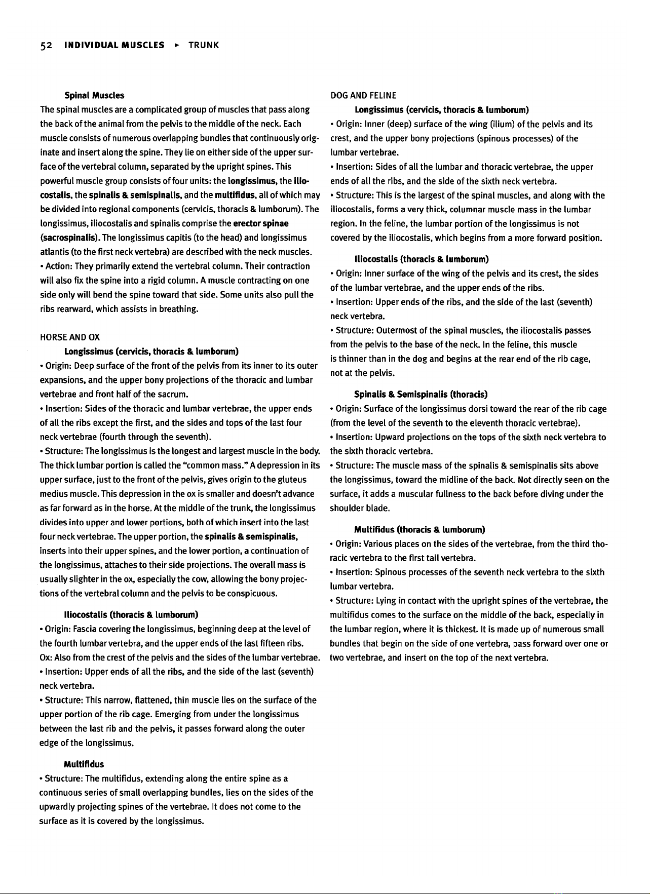
Spinal Muscles
The
spinal muscles
are a
complicated group
of
muscles that
pass
along
the
back
of the
animal from
the
pelvis
to the
middle
of the
neck.
Each
muscle
consists
of
numerous overlapping bundles that continuously
orig-
inate
and
insert
along
the
spine. They
lie on
either side
of the
upper sur-
face
of the
vertebral column, separated
by the
upright
spines. This
powerful muscle group consists
of
four
units:
the
longissimus,
the
ilio-
costalis,
the
spinalis
&
semispinalis,
and the
multifidus,
all of
which
may
be
divided
into
regional components
(cervicis,
thoracis
&
lumborum).The
longissimus,
iliocostalis
and
spinalis
comprise
the
erector spinae
(sacrospinalis).
The
longissimus
capitis
(to the
head)
and
longissimus
atlantis
(to the
first
neck vertebra)
are
described with
the
neck muscles.
•
Action: They primarily extend
the
vertebral column. Their contraction
will
also
fix the
spine
into
a
rigid
column.
A
muscle contracting
on one
side
only
will
bend
the
spine toward that side.
Some
units
also
pull
the
ribs
rearward, which assists
in
breathing.
HORSE
AND
OX
Longissimus
(cervicis, thoracis
&
lumborum)
•
Origin:
Deep
surface
of the
front
of the
pelvis from
its
inner
to its
outer
expansions,
and the
upper bony projections
of the
thoracic
and
lumbar
vertebrae
and
front half
of the
sacrum.
•
Insertion:
Sides
of the
thoracic
and
lumbar vertebrae,
the
upper ends
of all the
ribs
except
the
first,
and the
sides
and
tops
of the
last four
neck
vertebrae (fourth through
the
seventh).
•
Structure:
The
longissimus
is the
longest
and
largest muscle
in the
body.
The
thick
lumbar
portion
is
called
the
"common
mass."
A
depression
in its
upper surface, just
to the
front
of the
pelvis,
gives
origin
to the
gluteus
medius muscle. This depression
in the ox is
smaller
and
doesn't advance
as
far
forward
as in the
horse.
At the
middle
of the
trunk,
the
longissimus
divides
into
upper
and
lower
portions,
both
of
which insert into
the
last
four neck vertebrae.
The
upper
portion,
the
spinalis
&
semispinalis,
inserts into
their
upper spines,
and the
lower
portion,
a
continuation
of
the
longissimus,
attaches
to
their
side
projections.
The
overall
mass
is
usually
slighter
in the ox,
especially
the
cow,
allowing
the
bony projec-
tions
of the
vertebral column
and the
pelvis
to be
conspicuous.
Iliocostalis (thoracis
&
lumborum)
•
Origin:
Fascia
covering
the
longissimus,
beginning
deep
at the
level
of
the
fourth lumbar vertebra,
and the
upper ends
of the
last fifteen
ribs.
Ox:
Also from
the
crest
of the
pelvis
and the
sides
of the
lumbar vertebrae.
•
Insertion: Upper ends
of all the
ribs,
and the
side
of the
last
(seventh)
neck
vertebra.
•
Structure: This narrow, flattened,
thin
muscle
lies
on the
surface
of the
upper portion
of the rib
cage. Emerging from under
the
longissimus
between
the
last
rib and the
pelvis,
it
passes forward along
the
outer
edge
of the
longissimus.
Multifidus
•
Structure:
The
multifidus,
extending
along
the
entire spine
as a
continuous series
of
small
overlapping bundles,
lies
on the
sides
of the
upwardly projecting spines
of the
vertebrae.
It
does
not
come
to the
surface
as it is
covered
by the
longissimus.
DOG
AND
FELINE
Longissimus
(cervicis, thoracis
&
lumborum)
•
Origin:
Inner (deep) surface
of the
wing
(ilium)
of the
pelvis
and its
crest,
and the
upper bony projections (spinous processes)
of the
lumbar vertebrae.
•
Insertion: Sides
of all the
lumbar
and
thoracic vertebrae,
the
upper
ends
of all the
ribs,
and the
side
of the
sixth
neck vertebra.
•
Structure: This
is the
largest
of the
spinal muscles,
and
along
with
the
iliocostalis,
forms
a
very
thick,
columnar muscle mass
in the
lumbar
region.
In the
feline,
the
lumbar portion
of the
longissimus
is not
covered
by the
iliocostalis,
which begins from
a
more forward
position.
Iliocostalis (thoracis
&
lumborum)
•
Origin:
Inner surface
of the
wing
of the
pelvis
and its
crest,
the
sides
of the
lumbar vertebrae,
and the
upper ends
of the
ribs.
•
Insertion: Upper ends
of the
ribs,
and the
side
of the
last
(seventh)
neck
vertebra.
•
Structure: Outermost
of the
spinal muscles,
the
iliocostalis
passes
from
the
pelvis
to the
base
of the
neck.
In the
feline,
this
muscle
is
thinner than
in the dog and
begins
at the
rear
end of the rib
cage,
not
at the
pelvis.
Spinalis
&
Semispinalis (thoracis)
•
Origin:
Surface
of the
longissimus dorsi toward
the
rear
of the rib
cage
(from
the
level
of the
seventh
to the
eleventh thoracic vertebrae).
•
Insertion:
Upward projections
on the
tops
of the
sixth
neck vertebra
to
the
sixth thoracic vertebra.
•
Structure:
The
muscle mass
of the
spinalis
&
semispinalis
sits
above
the
longissimus,
toward
the
midline
of the
back.
Not
directly seen
on the
surface,
it
adds
a
muscular fullness
to the
back before
diving
under
the
shoulder blade.
Multifidus
(thoracis
&
lumborum)
•
Origin:
Various places
on the
sides
of the
vertebrae, from
the
third
tho-
racic
vertebra
to the
first
tail
vertebra.
•
Insertion: Spinous processes
of the
seventh neck vertebra
to the
sixth
lumbar vertebra.
•
Structure: Lying
in
contact with
the
upright
spines
of the
vertebrae,
the
multifidus
comes
to the
surface
on the
middle
of the
back, especially
in
the
lumbar region, where
it is
thickest.
It is
made
up of
numerous
small
bundles that begin
on the
side
of one
vertebra, pass forward over
one or
two
vertebrae,
and
insert
on the top of the
next vertebra.
INDIVIDUAL
MUSCLES
>
TRUNK
53
HORSE
DOG
54
INDIVIDUAL
MUSCLES
>
TRUNK
HORSE
OX
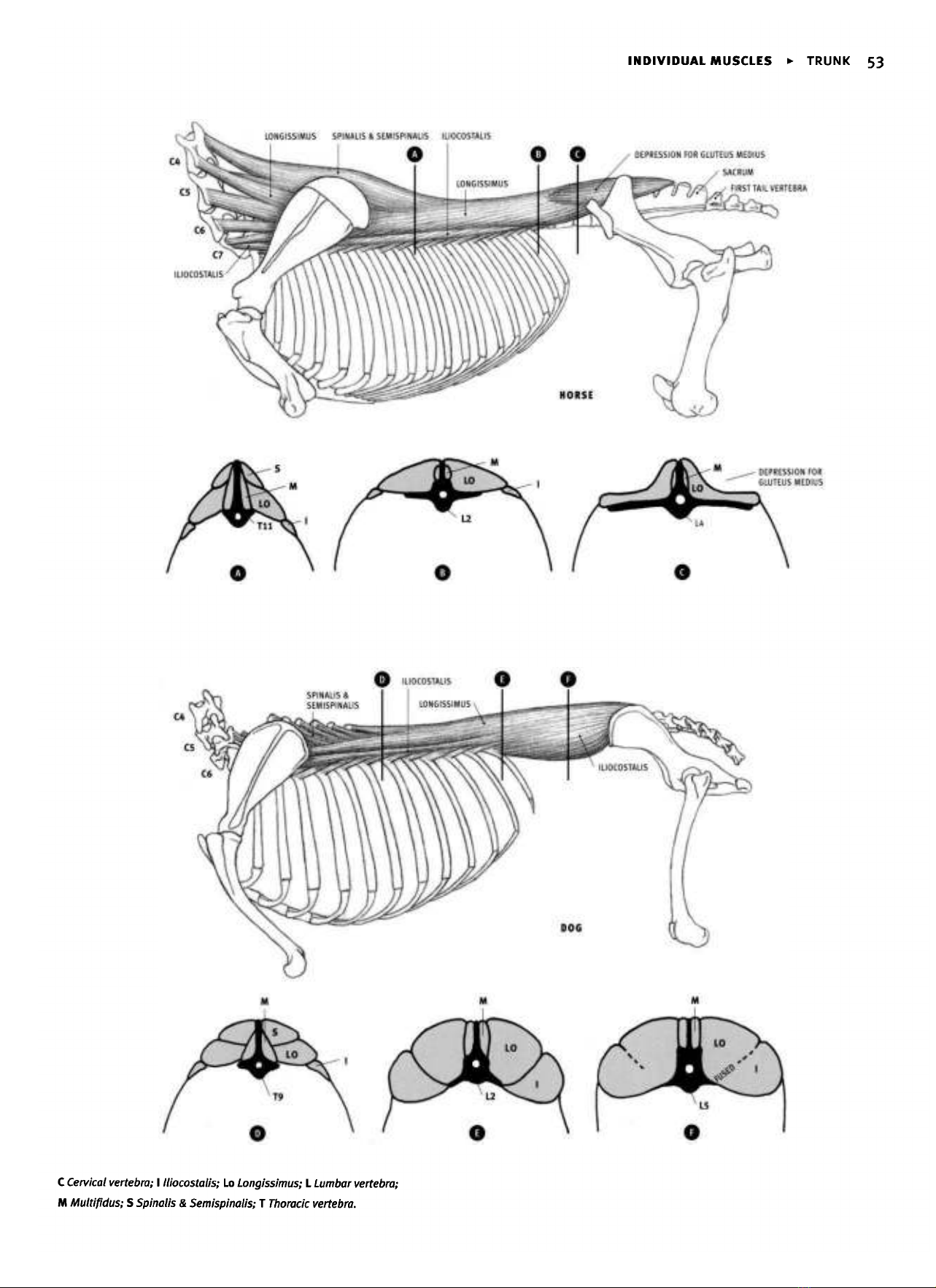
Internal abdominal oblique
(Obliquus
internus abdominis)
HORSE
•
Origin: Outer expansion
of the
front
of the
pelvis
("point
of the
hip")
•
Insertion: Inner
surface
of the
cartilage
of the
last four
or
five
ribs,
and
by
its
wide
tendon,
into
the
midline
on the
bottom
of the
abdomen
(linea
alba)
and the
front
end of the
bottom
of the
pelvis.
•
Action:
Compresses
the
abdomen
and
supports
its
contents; assists
in
bending
the
spine
to one
side.
•
Structure:
The
internal abdominal
oblique
is a
triangular,
fan-shaped
muscle
that develops
a
large,
wide tendon.
The
muscular portion
is
located
on the
upper portion
of the
side
of the
abdomen.
The
muscle
and
tendon
of
both sides
of the
body form
a
continuous
sling
that
pass-
es
under
the
abdomen
and
passively supports
the
abdominal contents
when
relaxed,
or
compresses them when
the
muscle
is
tensed.
The
wide
tendons from
each
side
of the
body fuse
on the
abdominal
midline,
contributing
to the
linea
alba.
The
linea alba
is a
tendinous thickening
of
the
midline
of the
abdomen that
passes
from
the
rear
end of the
sternum
to the
front
of the
bottom
of the
pelvis
(pubic
bone).
It is
formed
primarily
by the
fusion
of the
wide tendons
of
this
muscle
and
the
external abdominal
oblique.
OX
•
Origin: Also from
the
surface
of the
lumbar spinal muscle
(longissimus).
•
Insertion:
Most
of the
rear edge
of the
last
rib and its
cartilage,
and by
its
wide tendon
into
the
midline
on the
bottom
of the
abdomen
(linea
alba)
and the
front
end of the
bottom
of the
pelvis.
•
Structure: This muscle
is
irregular
in
shape rather than
triangular.
Muscle
fibers descending downward
and
forward from
the
point
of the
hip
form
a
raised
relief,
called
the
"cord
of the
flank." This ridge borders
the
rear side
of a
triangular depression,
the
"hollow
of the
flank."
The
lumbar spinal muscles border
the top of the
hollow,
and the
last
rib
defines
its
front
border.
The
cord
and the
hollow
are
usually
subtle
or
absent
in the
horse,
but
they
can be
quite prominent
in the ox,
with
the
cord
separating into
two or
three separate forms radiating from
the
point
of
the
hip. Muscle fibers
of
both
the
internal
and
external abdominal
obliques
are
present
in the
hollow,
filling
the
space
between
the rib
cage
and
the
pelvis.
This distance
is
greater
in the ox
than
in the
horse.
DOG
AND
FELINE
•
Origin: Side
of the
spinal muscle
in the
lumbar region; lower
end of the
crest
of the
ilium
at the
front
of the
pelvis.
•
Insertion:
Lower
end of the
last
rib and the
midline
of the
abdomen
via
the
wide tendon.
•
Structure:
The
internal abdominal oblique
lies
inconspicuously
on
the
side
of the
abdomen, mostly under cover
of the
external abdominal
oblique.
It
does
not
produce
the
cord
of the
flank
or the
hollow
of
the
flank.
INDIVIDUAL
MUSCLES
>
TRUNK
55
HORSE
DOG
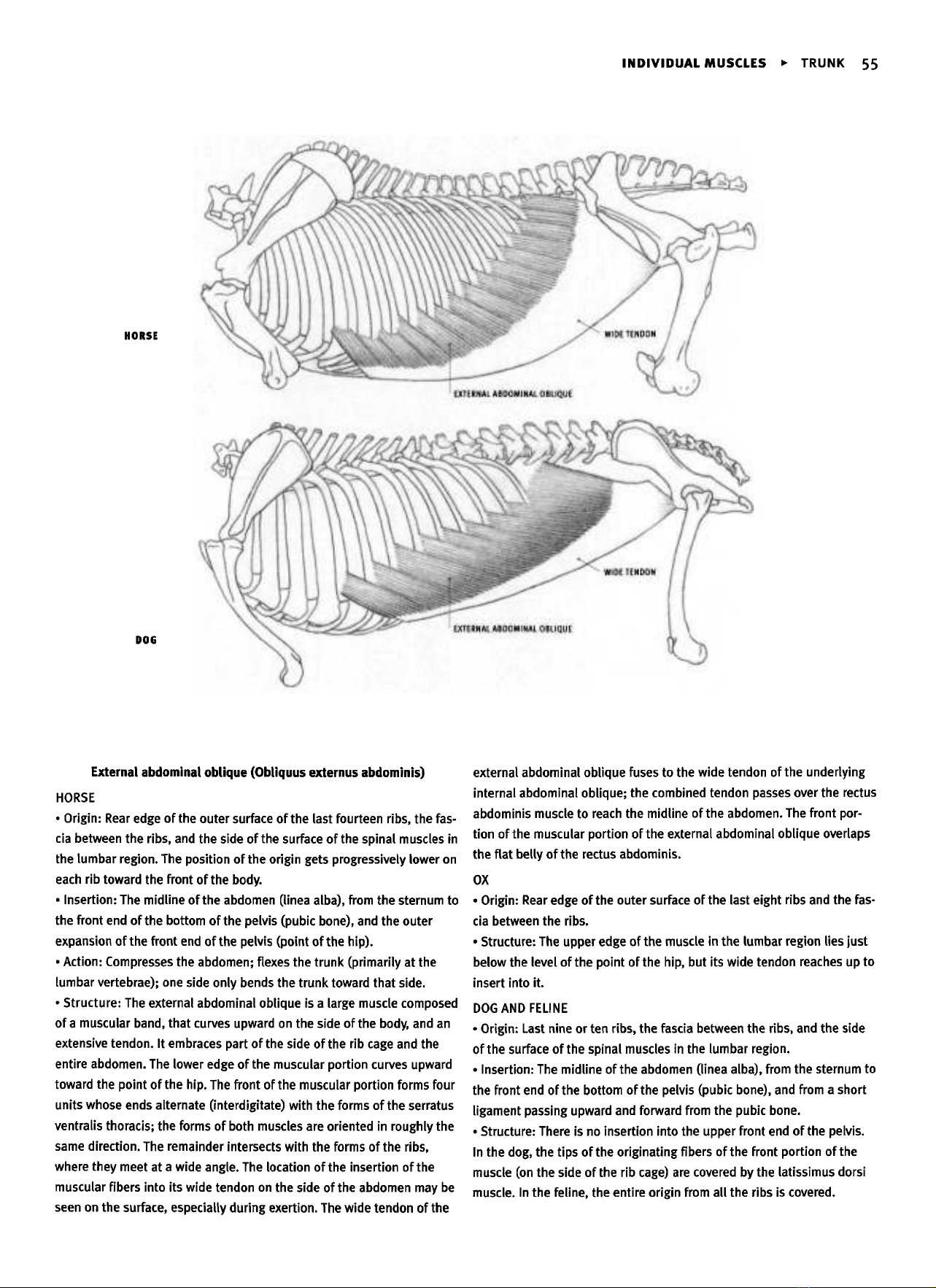
External
abdominal oblique (Obliquus externus abdominis)
HORSE
•
Origin:
Rear
edge
of the
outer surface
of the
last fourteen
ribs,
the
fas-
cia
between
the
ribs,
and the
side
of the
surface
of the
spinal muscles
in
the
lumbar region.
The
position
of the
origin
gets progressively lower
on
each
rib
toward
the
front
of the
body.
•
Insertion:
The
midline
of the
abdomen
(linea
alba),
from
the
sternum
to
the
front
end of the
bottom
of the
pelvis (pubic bone),
and the
outer
expansion
of the
front
end of the
pelvis
(point
of the
hip).
•
Action:
Compresses
the
abdomen; flexes
the
trunk
(primarily
at the
lumbar vertebrae);
one
side only bends
the
trunk toward that side.
•
Structure:
The
external abdominal
oblique
is a
large muscle composed
of a
muscular band, that curves upward
on the
side
of the
body,
and an
extensive
tendon.
It
embraces
part
of the
side
of the rib
cage
and the
entire abdomen.
The
lower edge
of the
muscular portion curves upward
toward
the
point
of the
hip.
The
front
of the
muscular portion forms four
units whose ends alternate
(interdigitate)
with
the
forms
of the
serratus
ventralis
thoracis;
the
forms
of
both muscles
are
oriented
in
roughly
the
same
direction.
The
remainder intersects with
the
forms
of the
ribs,
where
they meet
at a
wide
angle.
The
location
of the
insertion
of the
muscular
fibers
into
its
wide tendon
on the
side
of the
abdomen
may be
seen
on the
surface,
especially
during
exertion.
The
wide tendon
of the
external abdominal
oblique
fuses
to the
wide tendon
of the
underlying
internal abdominal
oblique;
the
combined tendon passes over
the
rectus
abdominis muscle
to
reach
the
midline
of the
abdomen.
The
front por-
tion
of the
muscular portion
of the
external abdominal oblique overlaps
the
flat
belly
of the
rectus
abdominis.
OX
•
Origin:
Rear
edge
of the
outer
surface
of the
last
eight
ribs
and the
fas-
cia
between
the
ribs.
•
Structure:
The
upper edge
of the
muscle
in the
lumbar
region
lies
just
below
the
level
of the
point
of the
hip,
but its
wide tendon
reaches
up to
insert into
it.
DOG
AND
FELINE
•
Origin:
Last
nine
or ten
ribs,
the
fascia between
the
ribs,
and the
side
of
the
surface
of the
spinal muscles
in the
lumbar region.
•
Insertion:
The
midline
of the
abdomen (linea
alba),
from
the
sternum
to
the
front
end of the
bottom
of the
pelvis (pubic bone),
and
from
a
short
ligament passing upward
and
forward from
the
pubic bone.
•
Structure:
There
is no
insertion into
the
upper front
end of the
pelvis.
In
the
dog,
the
tips
of the
originating
fibers
of the
front portion
of the
muscle
(on the
side
of the rib
cage)
are
covered
by the
latissimus dorsi
muscle.
In the
feline,
the
entire
origin
from
all the
ribs
is
covered.
56
INDIVIDUAL
MUSCLES
»
TRUNK
HORSE
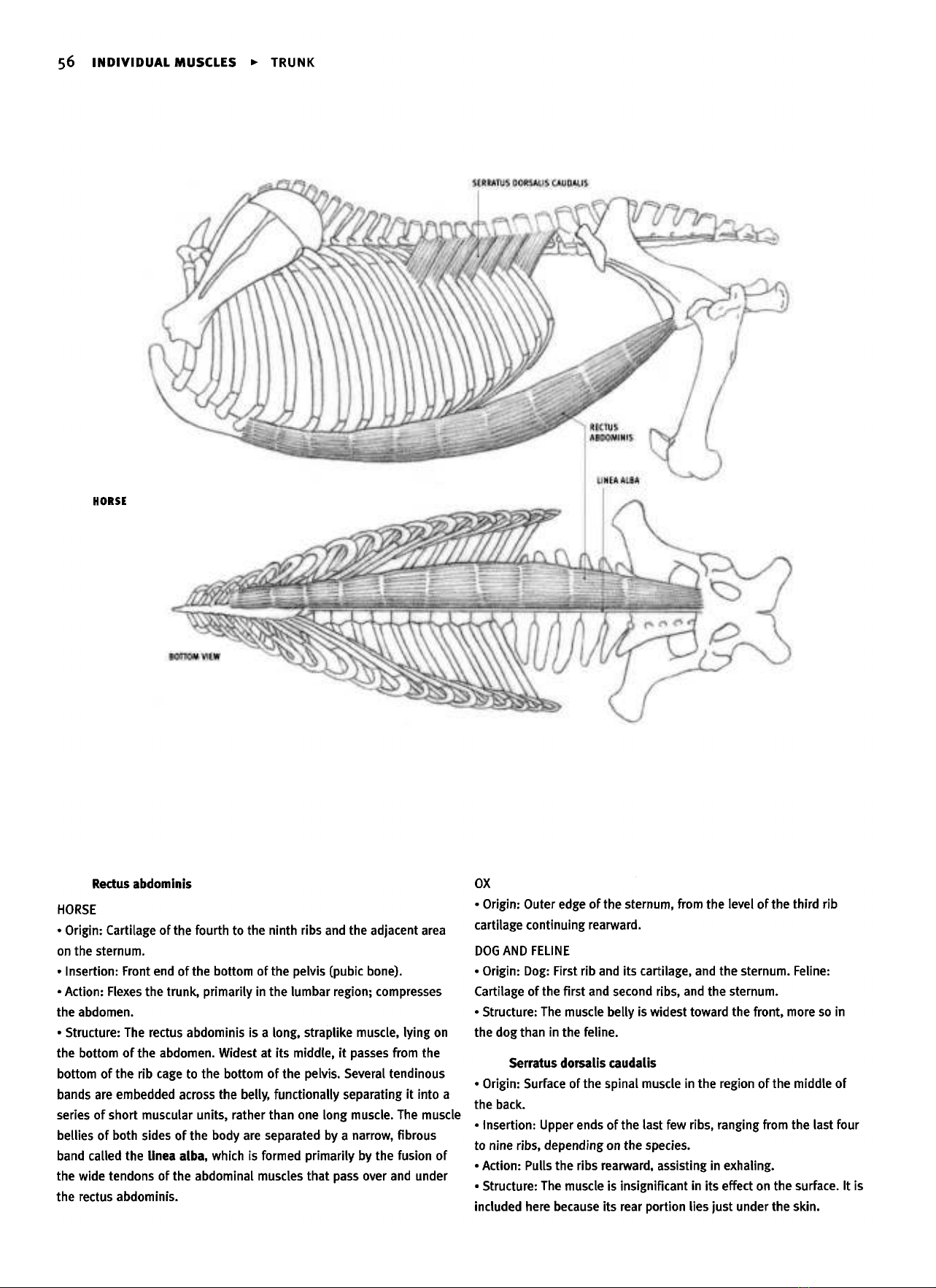
Rectus
abdominis
HORSE
•
Origin:
Cartilage
of the
fourth
to the
ninth ribs
and the
adjacent
area
on
the
sternum.
•
Insertion: Front
end of the
bottom
of the
pelvis (pubic bone).
•
Action: Flexes
the
trunk,
primarily
in the
lumbar
region;
compresses
the
abdomen.
•
Structure:
The
rectus abdominis
is a
long,
straplike muscle,
lying
on
the
bottom
of the
abdomen. Widest
at its
middle,
it
passes from
the
bottom
of the rib
cage
to the
bottom
of the
pelvis.
Several tendinous
bands
are
embedded across
the
belly,
functionally
separating
it
into
a
series
of
short muscular
units,
rather than
one
long
muscle.
The
muscle
bellies
of
both sides
of the
body
are
separated
by a
narrow, fibrous
band
called
the
linea
alba, which
is
formed primarily
by the
fusion
of
the
wide tendons
of the
abdominal muscles that pass over
and
under
the
rectus abdominis.
OX
•
Origin:
Outer edge
of the
sternum, from
the
level
of the
third
rib
cartilage continuing rearward.
DOG
AND
FELINE
•
Origin: Dog: First
rib and its
cartilage,
and the
sternum. Feline:
Cartilage
of the
first
and
second
ribs,
and the
sternum.
•
Structure:
The
muscle
belly
is
widest toward
the
front,
more
so in
the
dog
than
in the
feline.
Serratus
dorsalis caudalis
•
Origin:
Surface
of the
spinal muscle
in the
region
of the
middle
of
the
back.
•
Insertion: Upper ends
of the
last
few
ribs,
ranging from
the
last four
to
nine
ribs,
depending
on the
species.
•
Action: Pulls
the
ribs rearward, assisting
in
exhaling.
•
Structure:
The
muscle
is
insignificant
in its
effect
on the
surface.
It is
included here
because
its
rear portion
lies
just under
the
skin.